|
|
|
|
||||||||||||||||||||||||||||||||||||||||||||||||||||||||||||||||||||||||||||||||||||||||||||||||||||||||||||||||||||||||||||||||||||||||||||||||||||
|
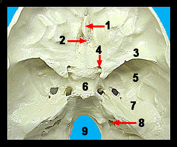
The sphenoid contains three pairs of processes: the greater wings, lesser wings, and the pterygoid processes. The superior surface of the sphenoid bone contains a saddle-like depression called the sella turcica that encloses the pituitary gland. Just superior and anterior to the sella turcica are the lesser wings of the sphenoid. Lateral to the sella turcica are the butterfly-shaped greater wings of the sphenoid bone. Notice the two optic foramina in the sphenoid bone just anterior to the sella turcica. The optic nerves from the eyes pass through these foramina. Immediately anterior to the sphenoid bone is the ethmoid bone, which forms the roof of the nasal cavity and a portion of the partition (nasal septum) dividing the nasal cavity into the right and left sides. The superior surface of the ethmoid bone forming the roof of the nasal cavity is called the cribriform plate. Note that this plate contains many tiny holes called olfactory foramina through which olfactory nerves pass from smell receptors in the nose to the brain. The crista galli projects superiorly from the midline of the cribriform plate. The dura mater anchors the brain to the cranial floor at this point of attachment. The perpendicular plate of the ethmoid extends inferiorly and forms the superior part of the nasal septum. |
|