|
|
|
|
||||||||||||||||||||||||||||||||||||||||||||||||||||||||||||||||||||||||||||||||||||||||||||||||||||||||||||||||||||||||||||||||||||||||||||||||||||||||||||||||||||||||||||||||||||||||||||||||||||||||||||||||||||||||||||||||||||||||||||||||||||||||||||||||||||||||||||
|
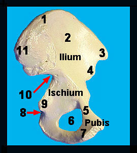
This image shows a medial view of the coxal (innominate) bone. The largest of the three bones making up the coxa is the ilium, which articulates with the sacrum at the roughened auricular surface. Immediately above the auricular surface is the iliac tuberosity. The prominent iliac crest runs anteriorly from the tuberosity to terminate in the anterior superior iliac spine. Anteriorly the superior ramus expands to form the pubic crest. The pubic crests from each coxal bone join anteriorly to form the pubic symphysis. An inferior ramus of the pubis comes off the pubic crest and fuses with the ischial ramus of the ischium, which articulates with the inferior ramus of the pubis. The fusion of the pubic and ischial rami results in the formation of a large foramen – the obturator foramen. Also note the ischial spine that projects medially from the ischium. Inferior to the ischial spine is the lesser sciatic notch through which a number of nerves and blood vessels pass to and from the pelvis and thigh. |
|