|
|
|
|
||||||||||||||||||||||||||||||||||||||||||||||||||||||||||||||||||||||||||||||||||||||||||||||||||||||||||||||||||||||||||||||||||||||||||||||||||||||||||||||||||||||||||||||||||||||||||||||||||||||||||||||||||||||||||||||||
|
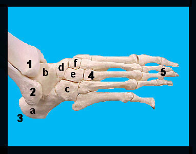
This image shows a medial view of the right foot. The foot bones are divided into the tarsals, metatarsals and phalanges (each of which is comprised of two or three separate bones). The seven tarsal bones (six of which are visible on this image) are larger and more specialized than the carpal bones of the wrist. The talus articulates with the fibula and tibia. The calcaneus ("heel bone") is the largest of the tarsal bones and is situated below the talus. The calcaneus helps support the weight of the body and serves for attachment of the large calf muscles. These calf muscles are attached to the calcaneus by the Achilles tendon. |
|