|
|
||||||||||||||||||||||||||||||||||||||||||||||||||||||||||||||||||||||||||||||||||||||||||||||||||||||||||||||||
|
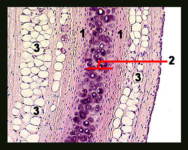
This slide shows a stained section of the trachea (windpipe). Note the rings of hyaline cartilage embedded in the walls of the trachea that provide support and help to maintain an open airway. Hyaline cartilage is the most common form of cartilage in the body, making up part of the nose, connecting ribs to the sternum and covering the articulating surfaces of bones. When sectioned and stained, the matrix of hyaline cartilage takes on a light purple color. Cartilage-forming cells called chondroblasts produce this matrix, which consists of an amorphous ground substance heavily invested with collagen fibers. Chondrocytes (mature cartilage cells) can be seen singly or in groups within spaces in the matrix called lacunae. The surface of all cartilage (except for articular cartilage) is covered by a membrane of connective tissue fibers called the perichondrium. Although the perichondrium is well-vascularized, cartilage tissue proper is avascular, which means that oxygen and nutrients have to diffuse from blood vessels in the perichondrium to the chondrocytes within the cartilage proper. |
|