|
|
|
|
||||||||||||||||||||||||||||||||||||||||||||||||||||||||||||||||||||||||||||
|
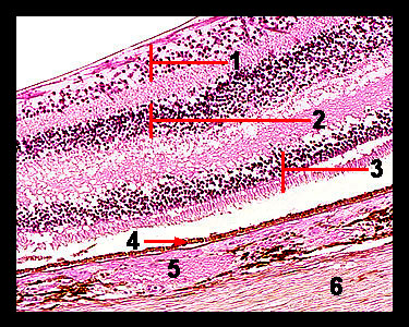
This slide shows a magnified view of the layers of the posterior portion of the eye. The outermost layer (which lies adjacent to the choroid) consists of a single layer of cells named the pigmented epithelium. The nervous layer of the retina is composed of three major cell types, photoreceptors (rods and cones), bipolar cells and ganglion cells. Although it is not possible to see where each of these three layers begins and ends, their dark, purple-staining nuclei are quite apparent. The first layer of rods and cones is found directly adjacent to the pigmented epithelial layer. Note that on the slide used for this photomicrograph, this layer has been torn away from the pigmented epithelium during preparation, leaving a white space of varying thickness between the two layers. The layer of rods and cones (which are the photosensitive cells of the eye) contains between 5-7 rows of prominent nuclei. The middle layer of bipolar cells also contains a number of rows of darkly staining nuclei. These cells conduct the electrical signals generated by the rods and cones to the ganglion cells of the third layer. The ganglion cell layer contains cells that generate an action potential and transmit it to the brain. The axons of the ganglion layer run across the inside layer of the retina to converge at the spot where they form the optic nerve. The spot where they pass through the retina to the outside of the eyeball as the optic nerve contains no rods or cones, so it is referred to as the blind spot or optic disc. |
|