|
|
||||||||||||||||||||||||||||||||||||||||||||||||||||||||||||||||||||||||||||||||||||||||||||||||||||||||||||||||||||||||||||||||||||||||||||||||||||||||||||||||||||||
|
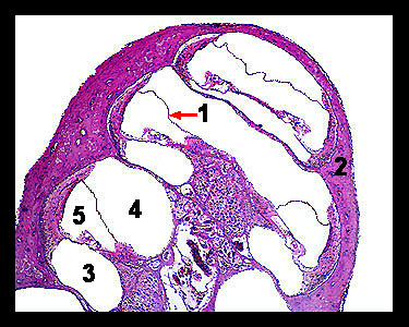
This slide shows a longitudinal section through a portion of the inner ear of a guinea pig called the cochlea (from the Greek word meaning "snail"), which consists of a coiled bony tube within which are three chambers that extend along its full length. The upper chamber, the vestibular duct, or scala vestibuli (so named because it is continuous with the vestibule), begins at the oval window of the middle ear. The lower passage is the tympanic duct, or scala tympani, which terminates at the membrane covered round window of the middle ear. Between these two chambers is a triangular section forming the cochlear duct (also known as the scala media). This duct is bounded on its upper surface by the vestibular membrane and on its lower surface by the basilar membrane. On the upper surface of the basilar membrane lies the organ of Corti, which contains the receptor cells of hearing. All these chambers contain fluid; perilymph within the scala tympani and scala vestibuli, and endolymph in the cochlear duct. |
|