|
|
||||||||||||
|
|
|
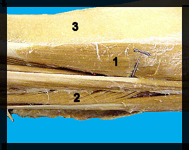
This image shows two muscles of the lateral surface of the lower leg of a male human cadaver. The most anterior of the muscles on the lateral surface of the shank is the tibialis anterior (cranialis). It originates from the lateral epicondyle and lateral surface of the tibia and it inserts on the first metatarsal and medial cuneiform. This muscle causes dorsiflexion in humans. The extensor digitorum longus is located just posterior to the tibialis anterior. It originates from the lateral condyle of the tibia and inserts on the dorsal surface of digits 2-5. This muscle produces dorsiflexion and toe extension in humans. |
|