|
|
||||||||||||||||||||||||||||||||||||||||||||||||||||||||||||||||||||||||||||||||||||||||||||||||||||||||||||||||||||||||||||||||||||||||||||||||||||||||||||||||||||||||||||||||||||||||||||||||||||||||||||||||||||||||||||||||||||||||||||||||||||||||||||||||||||||||||||||||||||
|
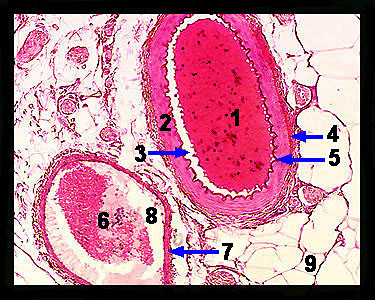
This image shows a cross section of an artery and a vein. Arteries are blood vessels that carry blood under high pressure from the heart (ventricles) to tissues. Veins are blood vessels that return blood under low pressure from the tissues to the heart (atria). Both arteries and veins are composed of three layers of tissue called tunics that surround a hollow core (lumen). The innermost layer (called the tunica intima, or interna) consists of a single layer of squamous, endothelial cells on top of a thin, elastic basement membrane called the internal elastic lamina. These endothelial cells form a smooth inner surface on the healthy lumen. The middle (usually much thicker) layer is the tunica media, which consists of layered smooth muscle cells along with elastic connective tissue fibers. These smooth muscle cells, which are under the control of the sympathetic nervous system, function to adjust the diameter of the lumen, thereby regulating the flow of blood through the vessel. The third, outermost layer is the tunica adventitia (externa), which is composed primarily of elastin and collagen connective tissue fibers, functions to support and protect blood vessels. Note that the wall of the artery is much thicker than that of the vein, reflecting the need to withstand the higher arterial pressures. For this reason, it may be difficult (as is the case in the image above) to distinguish the three layers of tissue in a vein. |
|