|
|
||||||||||||||||||||||||||||||||||||||||||||||||||||||||||||||||||||||||||||||||||||||||||||||||||||||||||||
|
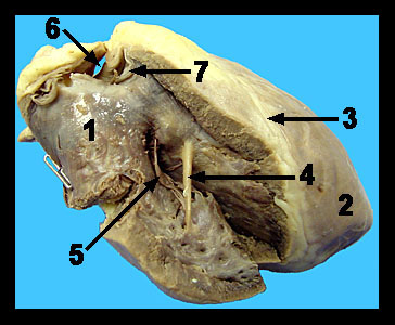
This image shows a close-up view of the inside of the right ventricle. Blood from the right atrium enters the right ventricle through the one-way tricuspid valve. The pulmonary trunk, carrying blood low in oxygen and high in carbon dioxide to the lungs, exits the right ventricle and curves toward the left side of the heart. Blood pumped into the pulmonary trunk is prevented from returning to the right ventricle by the pulmonary semilunar valve. A similar valve (the aortic semilunar valve) can be found at the base of the aorta where it exits the left ventricle. Each semilunar valve is composed of three pocket-like cusps with the pocket facing away from the ventricular chamber. When the ventricles contract during systole, the increasing blood pressure within the ventricles forces the cusps of the semilunar valves outward against the arterial walls so that the blood can pass into the aorta and pulmonary trunk. When the ventricles relax (diastole), the pressures within the ventricular chambers fall, and the back flow of arterial blood attempting to reenter the ventricles fills the pockets of the valves and snaps them shut. Notice the moderator band (septomarginal trabecula) running between the inner walls within the right ventricular chamber. As a modified form of one of the trabeculae carneae, the moderator band helps to carry signals to the papillary muscles, allowing them them to contract a few milliseconds before the rest of the right ventricle. |
|