|
|
||||||||||||||||||||||||||||||||||||||||||||||||||||||||||||||||||||||||||||||||||||||||||||||||||||||||||||||||
|
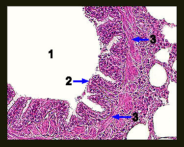
This image shows a cross section of a terminal bronchiole. Within the lung, tertiary bronchi become progressively smaller and eventually form small airways called terminal bronchioles. Note that this bronchiole is lined with a thick layer of ciliated columnar epithelium whose cilia help to remove foreign debris from the lungs. A thin (but complete) layer of smooth muscle that can regulate the diameter of the bronchiole can also be seen. |
|