|
|
||||||||||||||||||||||||||||||||||||||||||||||||||||||||||||||||||||||||||||||||||||||||||||||||
|
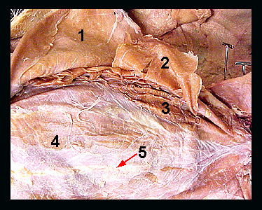
This image shows a ventral view of the abdominal wall of a cat, with the external oblique muscle reflected laterally. The abdominal wall consists of three sheet-like layers of muscle along with a band-like, longitudinal muscle located along the midventral line (linea alba). The linea alba is a thin, white longitudinal line of connective tissue that separates the right and left sides of the abdominal muscles. These muscles form a basket which encloses the viscera. Contraction of the abdominal muscles serves to compress the abdomen. The external oblique forms the most superficial of the three layers of abdominal muscles. This muscle originates from the posterior ribs and the lumbodorsal fascia. The fibers extend craniodorsally to insert along the length of the linea alba. Note that the muscle fibers of the external oblique do not extend all the way to the linea alba but are connected to it by an aponeurosis (a thin sheet of connective tissue). The internal oblique lies directly beneath the external oblique. Its fibers extend caudodorsally, nearly at right angles to the fibers of the external oblique. The internal oblique inserts by an aponeurosis on the linea alba. In the above image, the internal oblique has been cut and reflected to allow the transversus abdominis to be seen. The transversus abdominis originates from the posterior ribs and the transverse processes of the lumbar vertebrae. The fibers of this muscle extend ventrally and attach to the linea alba by an aponeurosis. Removal of the aponeuroses of the internal and external obliques will expose a longitudinal band of muscle lying lateral to the linea alba called the rectus abdominis. It originates from the pubis and inserts on costal cartilages 5-7 and the sternum. The rectus abdominis lies between the aponeuroses of the internal oblique and the transversus abdominis for most of its length. Contraction of the rectus abdominis compresses the abdomen and flexes the vertebral column. |
|