|
|
|||||||||
|
|
|||||||||||||||||||||||||||||||||||||||||||||||||||||||||||||||||||||||||||||||
|
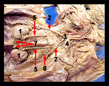
The urinary system of the female cat is essentially the same as in the male. Once again, observe the two bean-shaped kidneys located on the dorsal wall on either side of the vertebral column. Note the small adrenal glands, which resemble small lymph nodes in the cat. Draining each kidney is a tube called the ureter, which carries urine to the urinary bladder. A tube called the urethra conducts urine from the neck of the bladder to the outside of the body. Begin your observation of the reproductive system at ovaries, small oval-bodies located caudal to the kidneys. The lumpy appearance of the ovaries is due to the presence of Graafian follicles within the ovary. Each ovary is supported by a mesovarium, a mesenteric fold extending from a point near the caudal end of the kidney to the ovary and uterus. The uterus is a Y-shaped (bipartite) structure consisting of two uterine horns and a single body. Each wing of the Y forms the horn of the uterus (uterine horn). Kittens will develop in the uterine horns. The bipartite design of the feline uterus explains, in part, why large litter sizes are possible. The two uterine horns converge anterior to the pelvic canal and enter a common median passage. This would be the stem of the Y. The anterior part of this median passage is the body of the uterus. The body is not long and soon leads into the vagina, which proceeds posteriorly through the pelvic canal. The cervix separates the body of the uterus from the vagina. |
|