|
|
||||||||||||||
|
|
||||||||||||||||||||||||||||||||||||||||||||||||||||||||||||||||||||||||||||||||||||||||||||||||||||||||||||||||||||
|
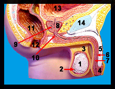
This image shows a model of a sagittal section of the human male reproductive system. The testes (the primary reproductive organs in the male) function to produce sperm and the male sex hormone testosterone. These structures are contained within a sac of skin called the scrotum that supports and protects the testes. Sperm are transported from the testes to the outside through a series of ducts. The first portion of the duct system is the highly coiled epididymis, which serves as a site for the storage and final maturation of sperm entering from its respective testis. The ductus deferens (also called the vas deferens) is a muscular tube that conducts sperm from the tail of the epididymis to the ejaculatory duct. The terminal portion of the ductus deferens that joins the ejaculatory duct is called the ampulla. The short ejaculatory duct is formed by a union of the ampulla of the ductus deferens and the duct of the seminal vesicle. The ejaculatory duct (which receives secretions from the seminal vesicle and prostate gland) penetrates the prostate gland where it empties into the urethra. The urethra serves as a common passageway (although not simultaneously) for both urine and semen. Note the seminal vesicle, bulbourethral gland and the prostate gland. These accessory glands produce the vast majority of semen (mixture of sperm and accessory gland secretions). The penis is composed mainly of erectile tissue and is a copulatory organ that delivers sperm into the female reproductive tract. The enlarged distal end of the organ is called the glans penis. Internally, the penis contains the penile urethra and three cylindrical bodies of erectile tissue. These three bodies consist of the paired corpora cavernosa and the corpus spongiosum that surrounds the urethra. The erectile tissue contains vascular spaces that become engorged with blood during sexual arousal. Also seen on the model are structures belonging to the digestive and urinary systems. |
|
