|
|
||||||||||||||
|
|
||||||||||||||||||||||||||||||||||||||||||||||||||||||||||||||||||||||||||||||||||||||||
|
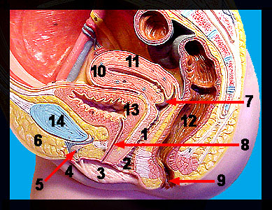
This image shows a model of a sagittal section of the human female reproductive system. The ovaries (seen on the next image) are the primary sex organs, and like those of the male, they produce both gametes (eggs) and sex hormones. Leading from the ovaries are the uterine tubes (oviducts), which receive an ovulated egg and serve as the normal sites of fertilization. The distal end of each uterine tube (infundibulum) is funnel-shaped and open-ended. The uterus (the site of prenatal development) is a thick walled, muscular organ with three anatomic regions: the fundus, body and cervix. The cervical canal connects the uterine cavity to the vagina, a thin-walled, muscular tube about 9 cm in length. The primary functions of the vagina are the reception of semen from the penis during intercourse and provision of a passageway for the delivery of a baby. The female reproductive structures located external to the vagina are called the external genitalia or vulva. These structures include the mons pubis, labia, vestibule and clitoris. The mons pubis is a subcutaneous pad of adipose tissue that covers the pubic symphysis region. The labia majora (two elongated skin folds that pass posteriorly from the mons pubis) are homologous to the scrotum in the male and serve to cover and protect other organs of the vulva. Beneath the labia majora are the labia minora, two smaller, thinner folds of skin that surround a recessed area called the vestibule. Within the vestibule are the external openings of the urethra (urethral orifice) and the vagina (vaginal orifice). Just anterior to the vestibule is a richly innervated, small, rounded structure composed of erectile tissue called the clitoris that is homologous to the penis. As in the previous models of the male reproductive system, this image also shows structures belonging to the digestive and urinary systems of the female. |
|
