|
|
|
|
||||||||||||||||||||||||||||||||||||||||||||||||||||||||||||||||||||||||||||
|
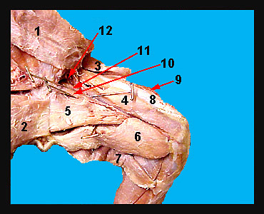
In the above image of the medial surface of the cat thigh, the sartorius and gracilis muscles have been cut and reflected back and the fascia lata removed to expose three of the four muscles which comprise the quadriceps group. These muscles pass to the knee joint and function to extend the shank. The large muscle on the anterio-lateral side of the thigh is the vastus lateralis. The muscle on the medial surface of the thigh is the vastus medialis. The rectus femoris lies on the front of the thigh between the vastus lateralis and the vastus medialis. The vastus muscles all originate on the femur and insert on the tibia via the patellar ligament. These muscles act across the knee joint to cause extension of the shank. In contrast, the rectus femoris originates on the ilium but still acts across the knee joint to extend the shank. At the posterior edge of the thigh is the semitendinosus. The large muscle anterior to the semitendinosus is the semimembranosus, which extends the thigh. The adductor femoris lies anterior to the semimembranosus. Both of these muscles originate on the pelvic girdle and insert on the femur. Two small, triangular shaped muscles lie anterior to the adductor femoris. The muscle adjacent to the adductor femoris is the adductor longus, and the most anterior muscle is the pectineus. Both the adductor longus and the pectineus arise from the pubis and insert near the proximal end of the femur. Both muscles adduct the thigh. The iliopsoas complex lies anterior to the pectineus. The largest muscle of this group is the psoas major and its action is to flex and rotate the thigh. |
|
