|
|
||||||||||||||||||||||||||||||||||||||||||||||||||||||||||||||||||||||||||||||||||||||||||||||||||||||||||||||||||||||||||||||||||||||||||||||||||||||||
|
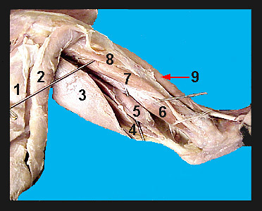
The large group of calf muscles on the posterior surface of the cat shank consists of the gastrocnemius, plantaris and soleus. The gastrocnemius is composed of a lateral and medial head. The origin of the lateral head of the gastrocnemius is the lateral epicondyle of the femur, lateral surface of the patella and adjacent tibia. The medial head originates from the medial epicondyle of the femur. The plantaris is sandwiched between the two heads of the gastrocnemius. The soleus arises from the proximal fibula. It is best seen from a lateral view. All three muscles converge to form the Achilles tendon which inserts on the calcaneus. In the cat, the action of these muscles is to cause extension of the foot. They are particularly important in pushing the foot off the ground. Their large size in the cat can account, in part, for the cat's great leaping ability. The triangular shaped popliteus originates from the lateral epicondyle of the femur and passes posterior to the knee joint to insert on the proximal end of the tibia. Its action is to flex the shank and rotate it slightly. The flexor digitorum longus is a long, thin muscle on the medial surface of the shank just posterior to the tibia. It originates from the head of the fibula and the shaft of the tibia. The flexor hallucis longus lies lateral to the flexor digitorum longus. Its origin is the shaft of the fibula and tibia. The tendons of these two muscles pass posterior to the medial malleolus. These two muscles flex the toes and foot. The small tibialis posterior (not clearly shown on the above image) lies beneath the flexor digitorum longus and between the flexor digitorum longus and flexor hallucis longus. It originates from the fibula and tibia and in part from the surface of the adjacent muscles. Its long, slender tendon passes posterior to the medial malleolus and inserts on ankle bones. Its job is extension of the foot. |
|
