|
|
||||||||||||||||||||||||||||||||||||||||||||||||||||||||||||||||||||||||||||||||||
|
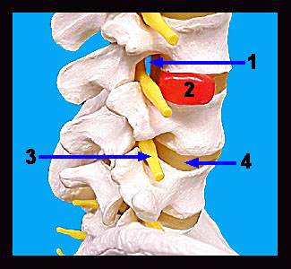
This image shows a model of the lower portion of the vertebral column. Note that the vertebrae are separated by discs of fibrous cartilage that cushion the bones from shock. Each disc is composed of an outer fibrocartilaginous ring called the annulus fibrosus and a soft, inner, highly elastic structure called the nucleus pulposus. Spinal nerves leave the spinal cord at regular intervals through the intervertebral foramina formed by the union of two vertebrae. As shock absorbers, the intervertebral discs are continually subjected to strong compressional forces. Occasionally, the discs between the lower vertebrae may become injured or weakened such that a portion of the nucleus pulposus protrudes into the intervertebral foramen, pressing on one of the spinal nerves in the process. This painful condition is called a herniated (slipped) disc. |
|