|
|
|
|
||||||||||||||||||||||||||||||||||||||||||||
|
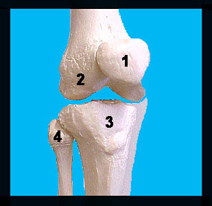
This image shows an anterior view of the bones that make up the knee joint. At the distal end of the femur, note the conspicuous lateral and medial condyles that articulate with the tibia of the leg to form a hinge joint that allows for flexion and extension at the knee. The smooth surface between the condyles on the anterior surface of the femur articulates with the patella (knee cap). The patella is a small, flat, triangular sesamoid bone enclosed in the tendon that secures the anterior thigh muscles to the tibia. It helps protect the knee joint anteriorly and improves the leverage of the thigh muscles acting on the joint. Also note the non-weight-bearing fibula on the lateral surface of the leg; the fibula does not articulate with the femur. |
|