|
|
||||||||||||||||||||||||||||||||||||||||||||||||||||||||||||||||||||||||
|
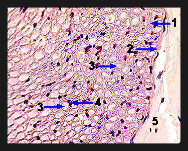
Note that in this magnified view of a nerve cross section that each individual nerve fiber consists of a tiny, centrally located axon surrounded by a larger Schwann cell sheath. In cross section, these nerve fibers look like small targets with the "bulls eye" being the axon! The empty space between these two structures would be filled in life with an insulating, fatty myelin sheath. Dark purple, crescent shaped Schwann cell nuclei can often be seen just outside of the Schwann cell sheaths. The spaces between individual nerve fibers are filled with a layer of loose connective tissue that constitutes the endoneurium. The nuclei of the fibroblasts responsible for producing this connective tissue can also be seen in the section. |
|