|
|
|
|
||||||||||||||||||||||||||||||||||||||||||||||||||||||||||||||||||||||||||
|
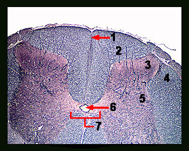
This image shows a magnified view of the posterior (dorsal) half of the spinal cord. Along the posterior median line of the spinal cord is a fissure called the posterior (dorsal) median sulcus. In the gray matter on the median lines lies a small central canal. This canal (filled with cerebrospinal fluid) runs the length of the spinal cord and is continuous with the ventricles of the brain. Notice that the gray matter is organized into posterior (dorsal) horns, which contain the nerve cell bodies of interneurons. Lateral horns of the gray matter containing nerve cell bodies of sympathetic preganglionic neurons are found in the thoracic and lumbar region of the spinal cord. The gray matter on each half of the spinal cord is joined together by the gray commissure, which contains the central canal. On each side of the spinal cord, the posterior horns of gray matter divide the white matter into columns called the posterior and lateral funiculi. These columns contain tracts made up of nerve fibers (axons) that carry action potentials to and from the brain and to other levels of the spinal cord. |
|