|
|
|
|
||||||||||||||||||||||||||||||||||||||||||||||||||||||
|
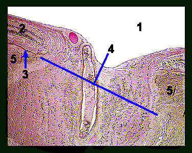
This slide shows a section through the optic nerve and the layers of the back of the eye. Note the optic nerve that leaves the back of the eye (where the blind spot is located) as well as the major layers of the eye. Observe the retina with its three layers of nuclei and thin dark layer of pigmented epithelium. Below the retina is the thin choroid, which contains dispersed patches of brown pigment and blood vessels. The outermost layer of the eye is the very thick, brown-staining sclera. |
|