|
|
||||||||||||||||||||||||||||||||||||||||||||||||||||||||||||||||||||||||||||||||||||||||||||||||||||||||||||||||||||||||||||||||||||||||||||||||||||||||||||||||||||||||||||||||||||||||||||||||||||||||||||||||||
|
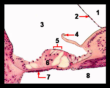
This magnified view of the cochlea shows the organ of Corti, which contains a complex arrangement of sensory receptor cells and membranes. The sensory cells of the organ of Corti are the columnar hair cells that have cilia on their exposed borders. Leading from the hair cells are nerve fibers that become part of the cochlear nerve. Forming a canopy over the hair cells is the gelatinous like tectorial membrane. The basilar membrane, which forms the floor of this organ, consists of tightly stretched fibers that are short near the vestibule and longer at the apex of the cochlea. Vibration of the basilar membrane displaces the hair cells, bending the hairlike projections, which generate nerve impulses in the afferent fibers connected to the cells. |
|