|
|
|
|
|
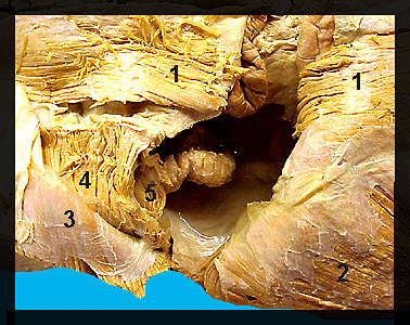
This image shows several of the abdominal muscles on a male human cadaver. The abdominal wall consists of three sheetlike layers of muscle along with a bandlike, longitudinal muscle located along the midventral line (linea alba). The linea alba is a thin, white longitudinal line of connective tissue that separates the right and left sides of the abdominal muscles. These muscles form a basket which encloses the viscera. Contraction of the abdominal muscles serves to compress the abdomen. The rectus abdominis originates from the pubis and inserts on costal cartilages 5-7 and the sternum. The external oblique forms the most superficial of the three layers of abdominal muscles. The internal oblique lies directly beneath the external oblique. Its fibers extend caudodorsally, nearly at right angles to the fibers of the external oblique. The transversus abdominis originates from the posterior ribs and the transverse processes of the lumbar vertebrae. |
|