|
|
|
|
||||||||||||||||||||||||||||
|
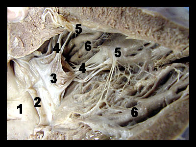
This image shows a close-up view of the inside of the left ventricle. Each side of the heart has a one-way valve between the atrium and the ventricle known as the atrioventricular valve. The atrioventricular (AV) valves of the two sides of the heart are similar in structure except that the right atrioventricular valve has three cusps while the left atrioventricular valve has two cusps. The right (AV) valve is termed the tricuspid valve while the left (AV) valve is termed the bicuspid valve. The bicuspid valve is also known as the mitral valve. Tendon-like strands of connective tissue known as chordae tendineae ("heart strings") run from the free edges of the cusps and attach on the ventricular wall to small projections of cardiac muscle called the papillary muscles. When the ventricles contract (systole), increased blood pressure within the chambers forces the cusps upward and together thereby closing the valve. This prevents the back flow of blood from the ventricle into the atrium. The chordae tendineae prevent the cusps from being pushed too far into the atria by the increased ventricular pressure. Muscular ridges called trabeculae carneae that project from the wall of the ventricle can also be seen in the image. |
|