|
|
||||||||||||||||||||||||||||||||||||||||||||||||||||||||||||||||||||||||||||||||||||||||||||||||||||||
|
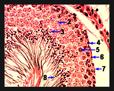
This image again shows a magnified view of a single seminiferous tubule in the testis. Observe the primary spermatocytes found just inside of the layer of spermatogonia. Primary spermatocytes will undergo the first meiotic division to produce two secondary spermatocytes (1n). The secondary spermatocyte is about half as large as the primary spermatocyte, and its nucleus contains diffuse, light-staining chromatin. The secondary spermatocyte very rapidly undergoes the second meiotic division to form spermatids (1n). There will be several layers of spermatids present in the wall of the seminiferous tubule. The young spermatids are very light in color and oval in shape. The spermatids become associated with Sertoli (sustentacular) cells and undergo a morphological transformation called spermiogenesis to become sperm. No genetic change during this transformation, but the cell loses its cytoplasm, forms an acrosomal cap and develops a flagellum. When spermiogenesis is complete, the sperm are released into the lumen of the tubules. The Sertoli cells (which are located against the basement membrane of the seminiferous tubule next to the spermatogonia) possess a large, round nucleus with distinct, black nucleolus. |
|