|
|
||||||||||||||||||||||||||||||||||||||||||||||||||||||||||||||||||||||||||||||||||||||||||||
|
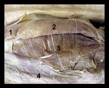
This image shows a ventral view of the abdominal wall of a cat, with the external oblique muscle reflected laterally. Note in this image the prominent transversus abdominis, which originates from the posterior ribs and the transverse processes of the lumbar vertebrae. The fibers of this muscle extend ventrally and attach to the linea alba by an aponeurosis. The rectus abdominis lies between the aponeuroses of the internal oblique and the transversus abdominis for most of its length. Contraction of the rectus abdominis compresses the abdomen and flexes the vertebral column. |
|