|
|
||||||||||||||||||||||||||||||||||||||||||||||||||||||||||||||||||||||||||||||||||||||||||||||||||||||||||||||||||||||||||||
|
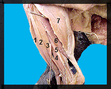
This image shows the muscles on the lateral side of the shank. The most anterior of the muscles on the lateral surface of the shank is the tibialis anterior (cranialis). It originates from the proximal end of the fibula and tibia and inserts on the first metatarsal. The action of this muscle is to flex the foot. The extensor digitorum longus is located just posterior to the tibialis anterior. It originates from the lateral epicondyle of the femur and inserts on the dorsal surface of specific phalanges. The extensor digitorum longus extends the digits. Three peroneus muscles lie posterior to the extensor digitorum longus and take their origin from the fibula. The peroneus longus is the most superficial of this group. It inserts on the proximal ends of all five metatarsals and its action is to flex the foot. The peroneus tertius is a tiny muscle lying directly beneath the peroneus longus. This muscle inserts on the first phalanx of the fifth digit. Its actions are to flex the foot and abduct and extend the fifth digit. The peroneus brevis lies posterior to the other peroneus muscles. It is the shortest of the three but has a relatively wide tendon. Its action is to extend the foot. The large group of calf muscles on the posterior surface of the shank consists of the gastrocnemius, plantaris and soleus . The gastrocnemius is composed of a lateral and medial head. The origin of the lateral head of the gastrocnemius is the lateral epicondyle of the femur, lateral surface of the patella and adjacent tibia. The medial head originates from the medial epicondyle of the femur. The soleus arises from the proximal fibula. It is best seen from a lateral view. All three muscles converge to form the Achilles tendon which inserts on the calcaneus. In the cat, the action of these muscles is to cause extension of the foot. They are particularly important in pushing the foot off the ground. Their large size in the cat can account, in part, for the cat's great leaping ability. |
|