Form & Function
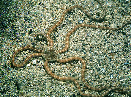
People often consider brittle stars and sea stars to be the same type of animal. They are very similar, however, brittle stars have arms that are longer and skinnier, and their tube feet do not have suckers on them like those of sea stars.
The brittle star, Ophiopsila riisei, has a disk diameter of about 12 millimeters. The disk has a gray color with black spots, with a blotchy appearance to it. This species is like other ophiocomids, in that they have both oral papillae and dental papillae (EOL 2003).
Ophiopsila riisei
can have up to to seven flat arm spines that are about 165 millimeters
(about 16 centimeters) long each. The arms of this brittle
 star are
irregularly banded with an off-white color and different shades of gray,
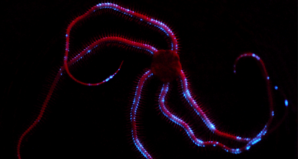
red, maroon, and purplish-brown (EOL 2003, Bocas 2003). These arms are
also unbranched and are not able to coil tightly, while other brittle
stars are capable of extreme coiling when other animals disturb them.
The arms of Ophiopsila riisei may coil slightly, but not nearly
as much as other species; their arms primarily move side to side (Pomory
2007). The image above shows the irregular banding and color of the arms
of Ophiopsila annulosa, a brittle star very closely related to
Ophiopsila riisei.
star are
irregularly banded with an off-white color and different shades of gray,
red, maroon, and purplish-brown (EOL 2003, Bocas 2003). These arms are
also unbranched and are not able to coil tightly, while other brittle
stars are capable of extreme coiling when other animals disturb them.
The arms of Ophiopsila riisei may coil slightly, but not nearly
as much as other species; their arms primarily move side to side (Pomory
2007). The image above shows the irregular banding and color of the arms
of Ophiopsila annulosa, a brittle star very closely related to
Ophiopsila riisei.
The tube feet of
an echinoderm are located on the oral surface of sea stars, and they
function in locomotion, respiration/gas-exchange. food gathering, and
sense-reception (Hajduk 1992, Mooi 1986). These feet are the exterior
distensions of the star's water-vascular system (Hajduk 1992).
Hemipholis elongata, a brittle star of the same class as
Ophiopsila riisei, has tube feet containing several layers. These
layers consist of: a ciliated epithelial layer lining the lumen of the
foot, a layer of longitudinal muscle, a connective tissue layer, and
finally a cuticle bilayer for a covering. The cells of the iner ciliated
epithelium layer could potentially have the job of circulating fluid in
the water-vascular system. The connective tissue layer is made of
collagen fibers arranged in longitudinal and circular directions;
nervous tissue components were found throughout the matrix of this layer
(Hajduk 1992). The outer epithelium layer of each tube foot is covered
by a thin cuticle layer, and has microvilli extending from its outer
surface (Hajduk 1992, Cobb and Moore 1986). These microvilli have been
observed and some seem to have been adapted for mucus secre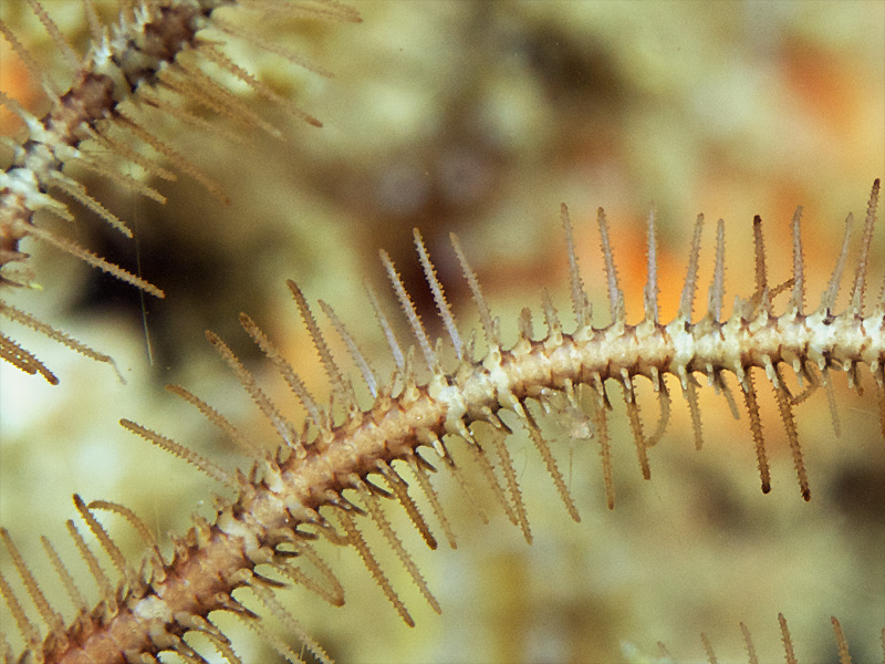 tion (Hajduk
1992).
tion (Hajduk
1992).
Retraction of each tube foot in ophiuroids is accomplished by contracting the longitudinal layer in the foot. When this muscular layer contracts, the fluid that fills the lumen of the tube foot is pushed into the tenacular extensions of the foot's wall. These extensions become limp and soft int he extended parts of the tube foot, and the appear enlarged or swollen in the contracted portion. The repition of this fluid transposition, although very little fluid is displaced, accounts for some of the locomotion of these brittle stars (Hajduk 1992).
For information about the reproduction process of Ophiopsila riisei, visit the Reproduction page!