|
|
|
|
||||||||||||||||||||||||||||||||||||||||||||||||||||||||||||||||||||||||||||||||||||||||||||||||||||||||||||||||||||||||||||||||||||||||||||||||||||
|
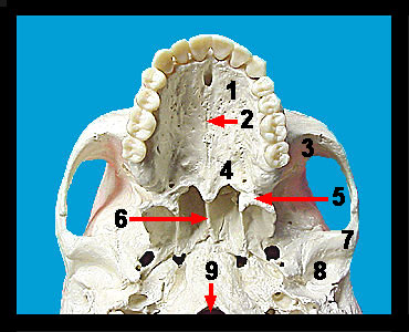
Immediately anterior to the temporal and inferior to the parietal bones and frontal bone is the butterfly-shaped sphenoid bone. The sphenoid has a very complex three-dimensional structure. In fact, parts of it can be seen in anterior, inferior, and posterior views of the skull as well as in the floor of the cranium. On the inferior surface of the skull the sphenoid bone forms a portion of the floor of the cranium and gives off the pterygoid processes, which serve as attachment sites for muscles important in chewing. The palatine processes of the maxillae form the anterior two-thirds of the hard palate which is the bony roof of the mouth. Note that they are joined together at the median palatine suture. If the palatine processes fail to grow together during early prenatal development, a cleft palate is produced. The maxillae house the upper teeth which are contained in sockets called alveoli within the alveolar margins. The palatine bones form the posterior third of the hard palate. The zygomatic bones form the cheekbones. They articulate with the zygomatic processes of the frontal and temporal bones as well as with zygomatic processes of the maxillae. The zygomatic bones also contribute to the orbits. |
||