|
|
||||||||||||||||||||||||||||||||||||||||||||||||||||||||||||||||||||||||||||||||||||||||||||||||||||||||||||||||||||||||||||||||||||||||||||||||||||||||||||||||||||||||||||||||||||||||||||||||||||||||||||||||||||||||||
|
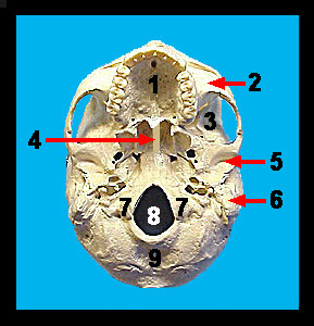
The occipital bone forms most of the posterior wall and base of the skull. This bone can best be seen in an inferior view of the skull. One of the most conspicuous features of this bone is the large opening (the foramen magnum) through which the spinal cord passes. On each side of the foramen magnum are the occipital condyles. These condyles are the smooth surfaces which articulate with the first cervical vertebra of the vertebral column. Also note the small oval mandibular fossa located on the inferior surface of the zygomatic process and just anterior to the external auditory meatus. The mandibular condyle of the mandible (lower jaw) fits into this fossa to form the temporomandibular joint. |
|