|
|
||||||||||||||||||||||||||||||||||||||||||||||||||||||||||||||||||||||||||||||||||||||||||||||||||||||||||||||||||||||||||||||||||||
|
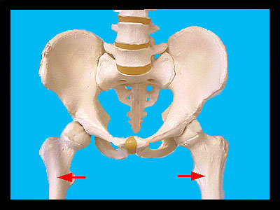
This image shows the proximal ends of the femurs (indicated by red arrows) as they are attached to the coxal bones of the pelvic girdle. The femur-coxal bone articulation (hip joint) is an example of a ball and socket joint. The ball (the head of the femur) articulates in the socket (the acetabulum) of the coxal bone. A ligament runs from the fovea capitis of the head to the acetabulum to secure this. The leg is moved by muscles much larger than those of the arm and this is reflected in the large processes of the femur. The neck of the femur separates the head from the large, rough greater trochanter. Medial and inferior to the greater trochanter is the lesser trochanter. The trochanters serve as attachment sites for muscles of the thighs and buttocks. The long, narrow diaphysis of the femur separates the proximal from the distal end. At the distal end of the femur the most conspicuous feature is the lateral and medial condyles. These condyles articulate with the tibia of the leg to form a hinge joint to allow for flexion and extension at the knee. On the posterior surface, the condyles are separated by the intercondyloid notch. The lateral and medial epicondyles are superior to the condyles and serve as attachment sites for muscles. |
|