|
|
|
|
||||||||||||||||||||||||||||||||||||||||
|
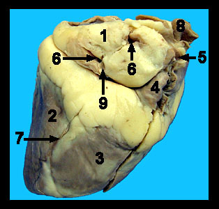
This image shows an external view of a preserved sheep heart. The heart is a four-chambered muscular pump. In adults it is approximately the size of a clenched fist. The four chambers consist of two thin-walled superior atria (singular atrium) and two thick-walled inferior ventricles. The heart is suspended in a double-walled fibroserous sac called the pericardium. The majority of this sac is usually absent from the prepared sheep hearts, but there may be parts of it still attached to the great vessels of the heart. The visceral layer of the pericardium, the epicardium, is attached to the outer surface of the heart wall. It is only one cell layer thick. |
|