|
|
|
|
||||||||||||||||||||||||||||
|
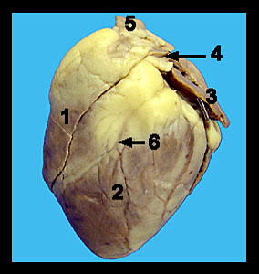
This image shows an external view of the anterior side of a preserved sheep heart. Note the pointed apex of the heart and the wide superior end of the heart which is termed the base. The large blood vessels (i.e., the great vessels of the heart) which carry blood to and from the heart are located at the base. The right and left atria are also located at the base and appear as thin-walled chambers with irregular, more or less scalloped edges. The wrinkled portion of each atrium that protrudes externally to form a pouch is called the auricle or atrial appendage. The atria serve as receiving chambers for low pressure venous blood returning to the heart thus their walls are extremely thin. Observe the anterior interventricular sulcus extending from the left side of the base obliquely to the heart's right side. The interventricular sulcus contains the left anterior descending coronary artery and the left coronary vein embedded within adipose tissue. The right ventricle lies to your left and toward the base relative to the anterior interventricular sulcus. The left ventricle lies to the right of the anterior interventricular sulcus and extends to and includes the apex of the heart. The ventricles are the pumping chambers of the heart and are, of necessity, thick walled. |
|