|
|
|
|
||||||||||||||||||||||||||||||||||||||||||||||||||||||||||||||||||||||||||||||||||||||||||||||||||||||||||||||||||||||||||||||||||||||||||||||||||||||||||||||||||||||||||||
|
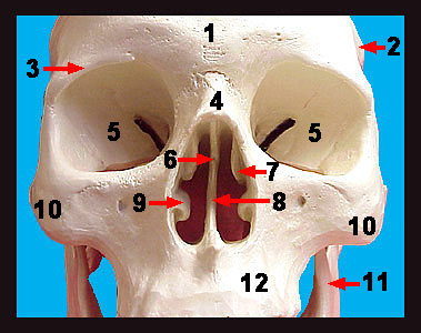
One of the most conspicuous facial features is the nose. However, the actual nasal bones supporting the nose are relatively small. The lacrimal bones form the anterior-most portion of the medial walls of the orbits. The inferior portion of the nasal septum and a portion of the floor of the nasal cavity is formed by the plough-shaped vomer bone. The maxillary bones, or maxillae (singular = maxilla) are the major bones of the face. They comprise a significant portion of the upper jaw and cheek bones of the individual. Notice that the maxillae also contribute to the orbits. The maxillae house the upper teeth which are contained in sockets called alveoli within the alveolar margins. An infraorbital foramen is located under each orbit. Nerves and blood vessels pass through this opening. The zygomatic process arises from the maxilla and articulates with the zygomatic bone. The coiled superior and middle nasal conchae (also called turbinates) extend medially from the lateral mass of the ethmoid. The paired inferior nasal conchae are separate bones (i.e., they are not part of the ethmoid bone) and form part of the lateral walls of the nasal cavity. Finally, note that the ethmoid forms most of the medial walls of the orbits. |
||