|
|
||||||||||||||||||||||||||||||||||||||||||||||||||||||||||||||||||||||||||||||||||||||||||||||||||||||||||||||||||||||||||
|
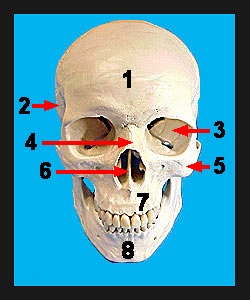
The skull is comprised of two main groups of bones, the cranial bones which form the cranium (a set of bones that forms the box enclosing the brain) and the facial bones (the bones comprising the anterior surface of the skull including the jaw). Except for the lower jawbone, all the bones of the skull are linked together by interlocking joints called sutures. Notice that there are numerous openings or "holes" in the skull. These openings are essential for nerves and blood vessels to enter and leave the cranium. These openings are termed foramina (singular, foramen). The frontal bone forms the anterior portion of the cranium commonly referred to as the forehead. Notice how this bone forms most of the roof of the orbit which houses the eyeball and immediately above the orbit, forms the supraorbital margin which lies directly beneath the eyebrows. The supraorbital margin is penetrated by the supraorbital foramen through which nerves and blood vessels pass. The smooth area between the orbits is called the glabella. Areas of the frontal bone just lateral to the glabella are filled with rather large cavities called the frontal sinuses. Superiolateral to the orbit a process of the frontal bone articulates with the zygomatic bone (cheekbone). This process is called the zygomatic process of the frontal bone. The frontal bone articulates posteriorly with the two parietal bones through the coronal suture. This suture derives its name from the fact it takes on the configuration of a crown sitting on the skull. |
|