|
|
|
|
|
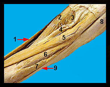
This image shows the anterior surface of the dissected thigh and hip of a human male cadaver. The longest muscle in the human body is the sartorius, which results in flexion of the thigh and weak flexion at the knee joint. Posterior to the sartorius, on the lateral surface of the thigh, is the tough and semitransparent fascia lata, which extends downward from a triangular muscle, the tensor fasciae latae. This muscle originates from the ilium and surface of adjacent muscles and inserts on the dorsal part of the iliotibial tract, and its contraction extends the shank and tightens the fascia lata. The anterior half of the thigh in the human contains the quadriceps group, which is involved primarily in the extension of the knee. Two of the muscles in this complex (the vastus lateralis and rectus femoris) are visible in the above image. The medial surface of the thigh, posterior to the sartorius and quadriceps group is covered by the gracilis, which adducts the thigh and flexes the leg. Its insertion is just inferior to the medial condyle of the tibia. Two small, triangular shaped muscles lie anterior to the adductor magnus. In humans, the adductor longus and pectineus are similar to those in the cat. The adductor longus functions to adduct and laterally rotate the thigh, while the pectineus adducts and flexes the thigh. |
|