|
|
|
|
|
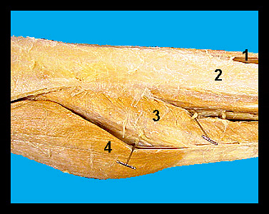
This image shows several of the principal muscles of the lower leg of a male human cadaver. The gastrocnemius is composed of a lateral and medial head. The origin of the lateral head of the gastrocnemius is the lateral epicondyle of the femur, lateral surface of the patella and adjacent tibia. The medial head originates from the medial epicondyle of the femur. The soleus arises from the proximal fibula. It is best seen from a lateral view. These muscles converge to form the Achilles tendon, which inserts on the calcaneus to cause extension (plantar flexion) of the foot. They are particularly important in pushing the foot off the ground. The most anterior of the muscles on the lateral surface of the shank is the tibialis anterior (cranialis). It originates from the proximal lateral epicondyle and the lateral surface of the tibia and it inserts on the first metatarsal and the medial cuneiform. This muscle causes dorsiflexion in humans. |
|