|
|
||||||||||||||||||||||||||||||||||||||||||||||||||||||||||||
|
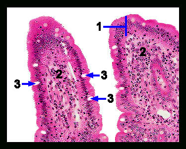
This image shows a magnified view of two villi. Note that each villus consists of a projection of the lamina propria covered by a layer of simple columnar epithelial cells, mucus-containing goblet cells and a scattering of enteroendocrine cells. Tiny projections of the plasma membrane of the epithelial cells of the mucosa called microvilli (difficult to see with a light microscope) further increase the absorptive surface area of each villus. It is across the epithelium-coated villi that nutrients, water and electrolytes are absorbed. |
|