|
|
||||||||||||||||||||||||||||||||||||||||||||||||||||||||||||||||||||||||||||||||||||||||||||
|
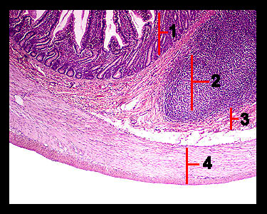
The histology of the ileum is similar to that of the duodenum except for the absence of Brunner's glands and appearance of more lymphatic tissue. Groups of lymphatic nodules (large enough to be seen with the naked eye) called Peyer's patches are present in the submucosa as well as in the mucosa. Each group consists of many nodules filled with dark staining lymphocytes. These structures extend to the lumen obliterating the villi in places. The increased concentration of lymphatic tissue toward the end of the small intestine reflects the fact that the large intestine contains huge numbers of bacteria that must be prevented from entering the blood stream. Note that there are fewer crypts of Lieberkühn in the ileum relative to the duodenum but goblet cells are much more plentiful. |
|