|
|
||||||||||||||||||||||||||||||||||||||||||||||||||||||||||||||||||||||||||||
|
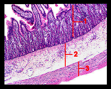
This image shows a cross section of the duodenum (the first portion of the small intestine). Once again, note the mucosa, submucosa, muscularis externa and adventitia that form the layers of the duodenal wall. Note: Since most of the duodenum is retroperitoneal, the outermost layer is called the adventitia. In general, the arrangement of the tissue layers in the small intestine is the same as that of the stomach. A noticeable difference is the presence of folds called plicae circulares and numerous finger-like villi that project into the lumen, slowing down the passage of food and providing a great increase in the total surface area for nutrient absorption. |
|