Interactions
Cyathocotyle bushiensis lives a parasitic lifestyle so it has created an interesting relationship with its multiple hosts. Its intermediate host, Bithynia tentaculata, does not suffer as severe affects as the definitive host suffers.

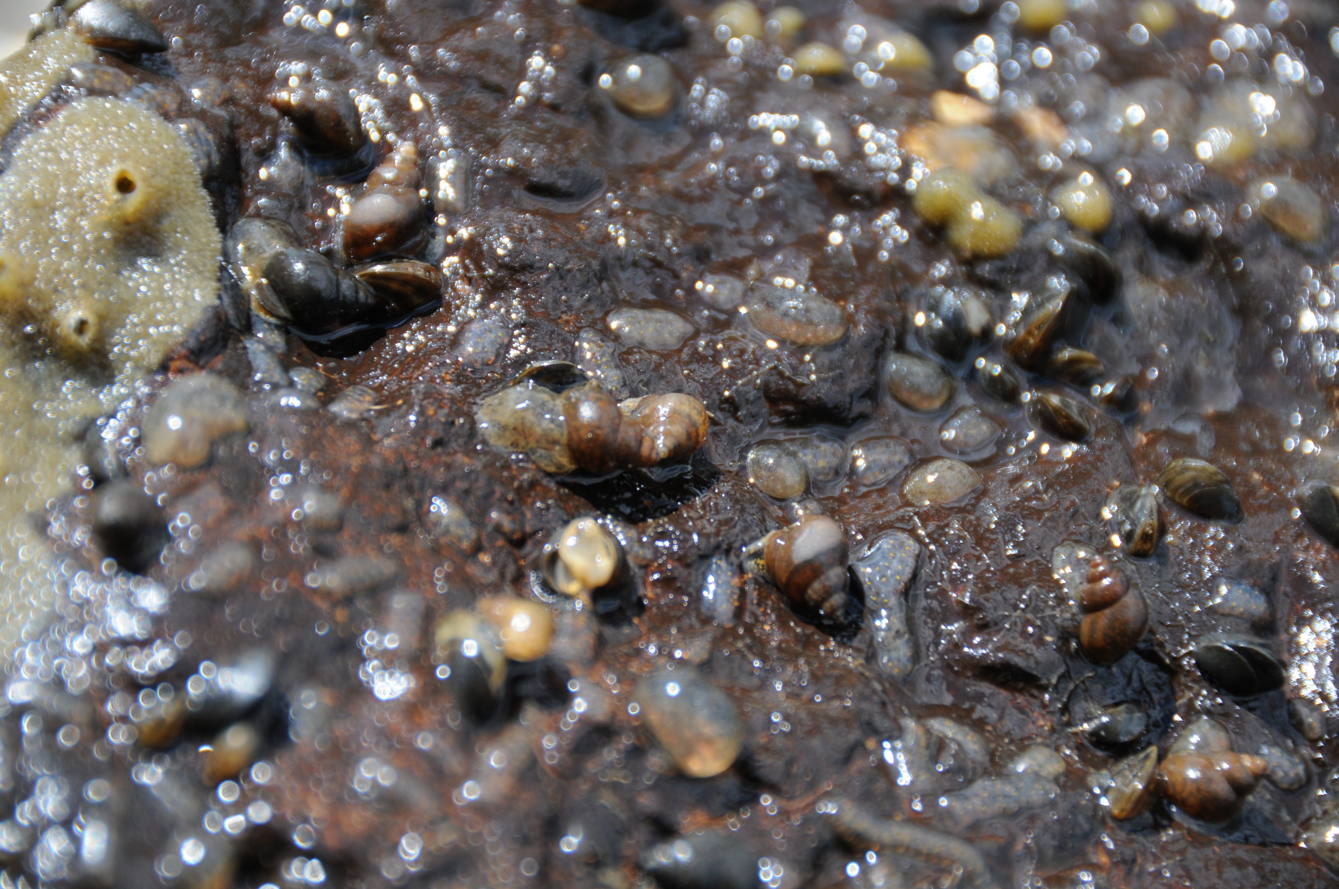
Above is a picture of B. tentaculata taken by Roger Haro.
B. tentaculata is where the parasite develops. According to Cheng in The Biology of Animal Parasites B. tentaculata is encysted by the metacercerial stage of C. bushiensis. The parasite will be found near the stomach of the snail, B. tentaculata or floating in the mantle cavity. Sometimes C. bushiensis can be found in the snail's intestine, which is near the gills. This does not occur very often (Gibson, Broughton, & Choquette, 1962).
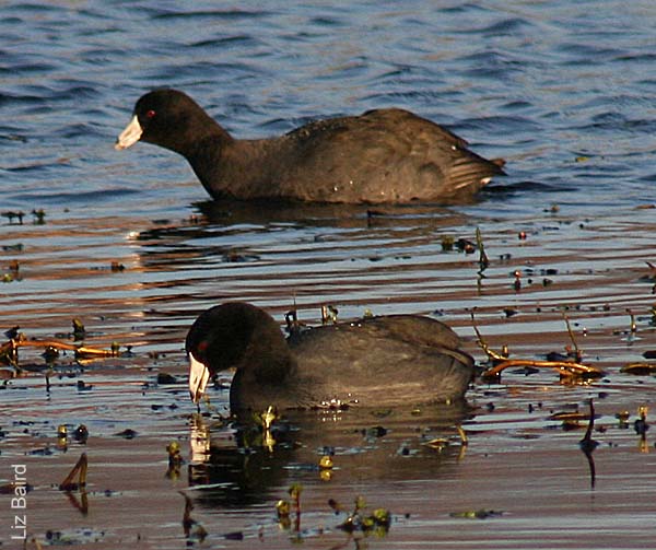
Following the infection of B. tentaculata a waterfowl bird (either Anas rubripes, Anas discors, or Anas carolinesis) ingest a snail. If B. tentaculata is infected the C. bushiensis will be passed to the waterfowl. After three to five days C. bushiensis has fully developed within the definitive host and begins to have an affect on the waterfowl's body and systems(Gibson, Broughton, & Choquette, 1962).
Inflammation of the caecum, known as typhlitis, is caused by
reactions to multiple problems caused by C. bushiensis (Gibson,
Broughton, & Choquette, 1962). There are three pathologies of the caeca that are
measured for the effects on the definitive host.
1. Hemorrhagic spots are where the parasite was attached to the caecal wall.
After a period of time C. bushiensis moves from one attachment point to
a new point of attachment, leaving behind a hemorrhagic spot. These spots are
made because C. bushiensis is feeding on the epithelial lining of the
intestine. It digests these tissues outside of its body, extracorporeally, and
leaves behind a hemorrhagic spot when it moves to an area where the epithelial
lining is still intact and begins to feed there (Hoeve & Scott, 1988).
2. When C. bushiensis is feeding it also causes blood and fibrin to
leak into the caecum. This is how plaques and cores are formed. They are caused
when the host responds to the presence of the parasite and the strength of the
response. The plaques are formed after C. bushiensis has left behind a
hemorrhagic spot. The immune response is delayed and finally takes affect after
the parasite has caused damage to the epithelial lining (Hoeve & Scott, 1988).
The waterfowl that did not have hemorrhagic spots had ulcerations of the caecal wall as well as generalized areas of dead cells. Some of the waterfowl also had the development of adhesions, which took place outside of the small intestine and caecal wall. These adhesions were between two of their caeca or between their small intestine and caeca. Another problem that could be seen from outside the lining of the caecal wall were breaks in the wall (Gibson, Broughton, & Choquette, 1962).
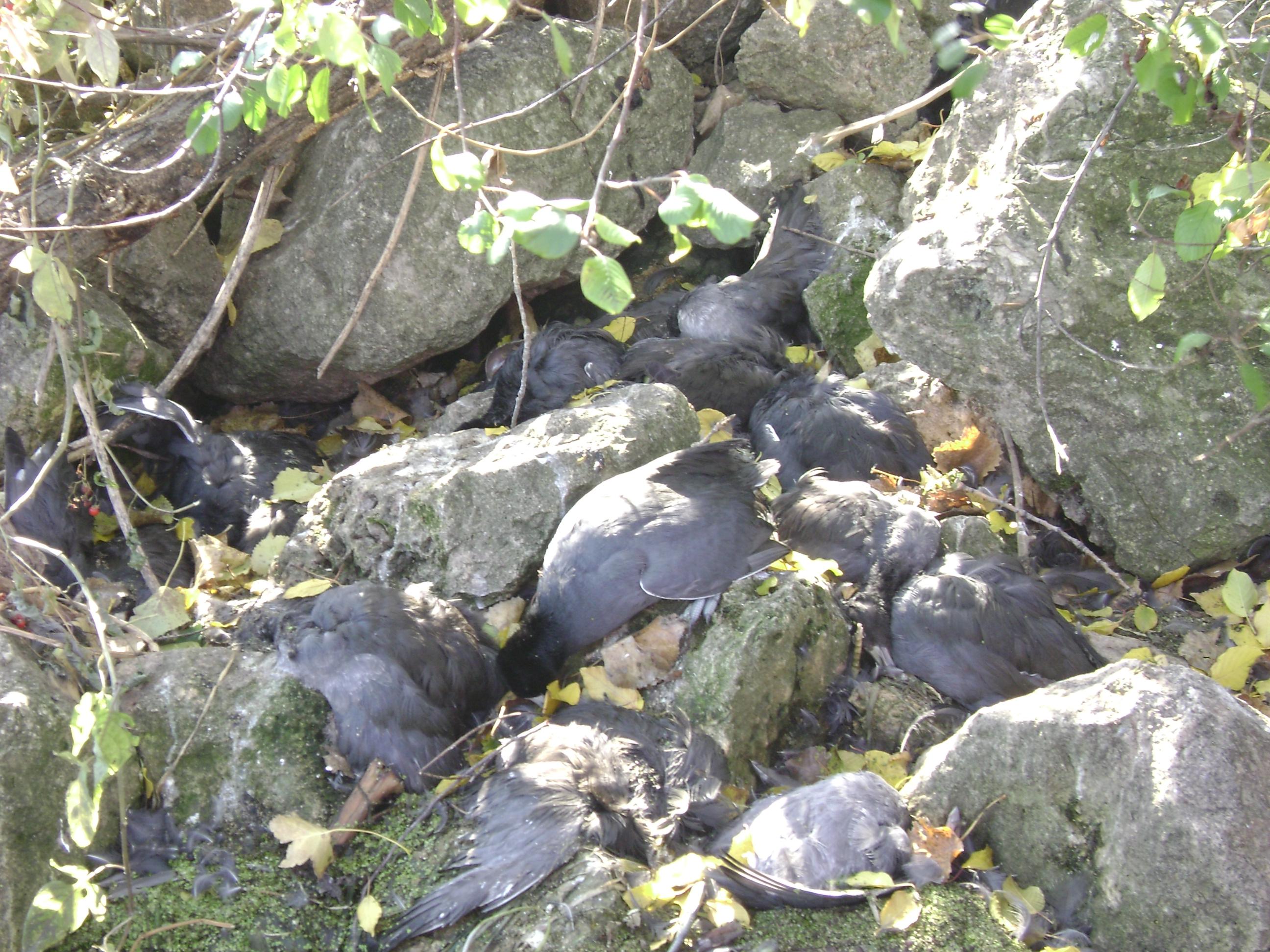
All of these problems, caused by C. bushiensis, resulted in the die-offs of large numbers of waterfowl (McDonald, 1981). These epizootics continue to occur where there is a presence of the intermediate host, B. tentaculata.

Above is a picture of dead coot. They died where they were sitting in the rocks.
Taken by Greg Sandland.
To learn more about the die-offs and why they occur see the Nutrition or Adaptation pages
