Echinococcus granulosus
A Parasitic Tapeworm
Cystic Hydatid Disease
Cystic Hydatid Disease, also known as echinococcosis, is a disease caused by the infection of a host with either the larval or adult form of the tapeworm of the genus Echinococcus. The cycle of infection of this parasite includes the infection of both a definitive host and an intermediate host by the different stages of the life cycle. The larval form occurs in the intermediate host, usually an herbivore such as deer, sheep and cattle. Typically, the definitive host is a carnivore, such as a dog or a wolf, who house the adult form of the parasite. Different species of the Echinococcus genus interact with their hosts slightly different; this site focuses primarily E. granulosus.
The adult tapeworm is usually very
small and resides in small intestine of the definitive host.
The eggs of the adult are excreted through the feces
of the definitive host. The eggs survive
longest in damp, cool places within the environment, but can
survive up to several years due to their highly resistant
nature. After being excreted from the
definitive host they are accidentally ingested by the
intermediate host via contaminated vegetation.
Once in the intermediate host, the hooked embryo
hatches and burrows into the small intestine of the host
organism. Here, the parasite enters the
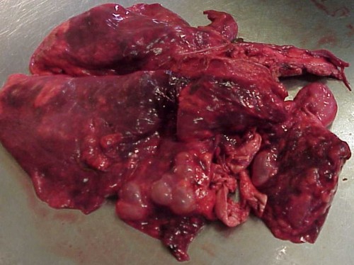
blood stream and eventually becomes lodged in an organ, most
commonly, the lungs. However,
Echinococcus can be found in the liver, kidney, brain,
bone marrow, skeletal muscle and the eye.
When humans are the intermediate host, the most common
location of infection is the liver. When
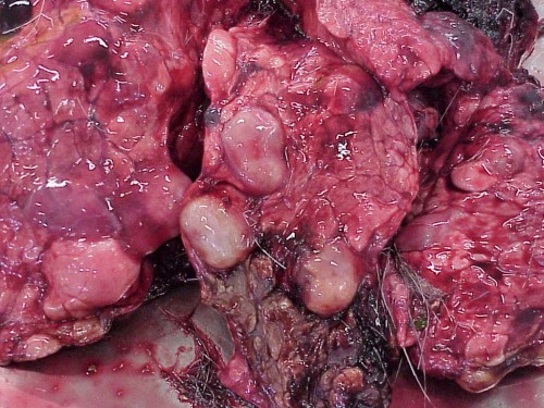
the larva reaches the target organ, it forms the hydatid
cyst. Within this cyst asexual budding
takes place to produce thousands of larval tapeworms.
The cycle is perpetuated once a carnivore ingested
the infected intermediate host, becoming the new definitive
host.
Typically infection of the definitive host, by the adult form of the parasite is asymptomatic and non pathogenic. However, the pathology of infection with the larval stage varies depending on the size, number and location of the cysts. Due to the pressure produced by the cyst, changes in the tissue can occur in the host. The most severe infections occur when the larva are located in the kidney, spleen, or brain tissue sometimes resulting in death of the host.
The diagnosis of the definitive host can be accomplished shortly after infection by using a test called Enzyme Linked Immunosorbent Assay (ELISA) to identify antigens of the parasite in the feces of the host. Diagnosis of the larval form of E. granulosus can be accomplished by identifying the presence of cysts in the infected organ.
Treatment of the definitive host can be accomplished with the administration of drugs such as Praziquantel or Arecoline. Arecoline works by paralyzing the parasite causing it to loosen its grip on the small intestine of the host; it can then be expelled via defecation. Treatment of the intermediate host is typically not necessary because most infections do not produce significant pathological damage and mortality is not usually affected. However, more severe infections, especially in humans are regularly treated. Humans are treated by surgical removal of the cyst from the infected organ. This is usually followed by the administration of different drugs to make sure all larval forms of the parasite are removed or killed. However, treatment is not always possible depending on severity of infection and the location of the cyst.
Now that you are an expert on E. granulosus, come learn a little bit about this site's author.
