
Anatomy and Physiology:
All spiders have a very similar body plan, and
when it comes to Phoneutria fera, things don’t
differ too much. They are all broken up into two
main segments; they include the prosoma or the
cephalothorax, and the opisthosoma or the
abdomen. These two segments are held together by
a structure called the pedicle. The prosoma is
the “head” region of the spider and contains all
eight legs, the eyes, the chelicera, pedipalps
and others. The opisthosoma then contains the
spinnerets, anal opening, “lungs”, heart, and
reproductive organs. This page will break each
section down both internally and externally as
well.
Cephalothorax:
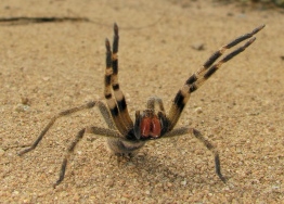
External: Perhaps the most
obvious external feature on the cephalothorax
are the eight jointed appendages or legs.
The big powerful legs which are moved with
striated skeletal muscles are the sole means of
locomotion for all spiders. These legs are
jointed in either four or five joints which all
allow for movement only in one plane, similar to
that of an elbow. The joint that connects the
legs to the cephalothorax allow for circular
movement similar to that of a shoulder or hip
joint. The first and fourth legs will be the
longer of the four and will be almost identical
in their anatomy whereas the second and third
legs will be shorter and be identical as well. The legs are also covered in
what appear to be very short hairs. These hairs
are actually receptors that are very sensitive
and can even sense changes in air pressure. This
will help them find a butterfly fluttering
through the air or help them avoid the buzzing
wings of their one main predator: The Tarantula
Hawks.
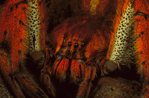
The next external parts of the cephalothorax
are the chelicerae. The
chelicerae are the
first appendages on the prosoma region and on
P.
fera are a noticeable red color. The chelicerae
are found right under the eyes and are composed
of two parts: the basal stout portion, and then
the movable fangs which are located underneath.
The fangs are used for
injecting venom or injuring prey, cutting their
silk threads (which is why the underside of the
fangs are serrated), and even grasping objects,
hence the chelicerae sometimes being referred to
as the spider’s hands.
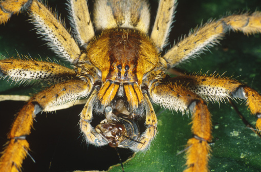
Inside or underneath the
chelicerae lie the Pedipalps or
mouth parts. These are the second set of
appendages and serve two main purposes. The
first is to serve as copulatory organs and are
used in the mating rituals of many spiders and
the second is to move and manipulate the prey of
the spider as they are eating it.
Internal:
Although the Eyes can be seen
externally, most of the eye lies beneath the
exoskeleton. P. fera has eight eyes arranged in
three rows. The first row has two(which are the
most functional and useful for the spider), the
second row has four, and the third row has two
eyes that are widely spaced from each other.
Each eye is connected to the main cephalized
ganglion, or “brain”, which can be found on the
ventral side of the cephalothorax.
The first
part of the Intestine is also
found within the cephalothorax and extends from
the mouth to through the pedicle into the
abdomen. The main muscle found within the head
region is the levator, or
pharynx muscle, which controls the mouth parts
of the spider. As stated before there exists a
main ganglion found internally
on the ventral side of the spider that serves as
its brain.
Pedicle: this term
simply refers to the thin waist like region of
the spider that attaches the cephalothorax to
the abdomen. This structure contains the aorta
connecting the heart in the abdomen to the
cephalothorax, a large nerve that connects the
two segments, and the intestine which carries
food brought in through the mouth on the
cephalothorax to the midgut which is in the
abdomen.
Abdomen:
External: The abdomen is fairly
limited in regards to its external anatomy. The
main structures that are found externally on the
abdomen are the spinnerets
which are used for spinning the web and are
located right below the anal opening. There are
three pairs of spinnerets which are all highly
coordinated because of the need for them to work
individually as well as with one another to
successfully spin their thread.
Above the
spinnerets is the anal opening
which is used to secrete digested waste. The
final external feature on the abdomen is the
reproductive opening which is
located on the ventral side of the spider. In
males this is where the sperm will leave the
body to fertilize the females. In females this
is where the sperm will enter and fertilize the
eggs and also where the fertilized eggs will
leave the body.
Internal: The abdomen houses a
variety of vital organs to the spider. The
heart lies dorsally along the
length of the abdomen. Spiders have a special
form of heart known as a tube heart which is
just as it is named. A muscular tube that helps
to pump the circulatory fluid throughout the
spider. Spiders have what is
known as a open circulatory system which is
characterized by a lack of closed veins and
arteries. When the heart beats the circulatory
fluid known as hemolymph is pushed through the
arteries and veins into the various sinuses of
the body.
Located on the ventral side of the
heart is the Midgut and
Malpighian Tubules.
The midgut brings in food from the intestinal
tract and is attached to several malpighian
tubules. The malpighian tubules brings the nutrients the spider needs from the midgut to
the various parts of the spider which need these
nutrients.
nutrients the spider needs from the midgut to
the various parts of the spider which need these
nutrients.
The main means of respiration are
also found internally on the abdomen and are
referred to as the
Book Lungs.
These "lungs" differ greatly from those found in
mammals. These lungs are found on the ventral
side of the spider and look similar to a book by
how they appear to be “pages” of air pockets
which are used for gas exchange and to oxygenate
the hemolymph that the atmospheric oxygen comes
in contact with. The book lungs are connected to
the heart so that the new oxygen rich hemolymph
can immediately be pumped throughout the body.
Book lungs more resemble gills than they
actually do lungs.
The final major abdominal internal structures
are the spinning glands which
are responsible for making the material that is
used to spin webs which connected to and
excreted out of the spinnerets.
Venom: The venom
utilized by P. fera is widely considered as the
deadliest in the world however certain
components of that same venom are being
researched for their use in the pharmaceutical
field. First we’ll discuss the deadly part of
the venom known as PhTx3. This
toxin is a deadly neurotoxin that inhibits
calcium ion channels in the nervous system
leading to paralysis, but also has a powerful
stimulating effect on serotonin receptors on
nerves throughout the body which
 causes a great
amount of pain. PhTx3 is known as a
broad-spectrum calcium
ion channel blocker which
interferes with the part of the nervous system
that is responsible for muscle contraction. If
enough of the venom is injected into the victim
full on paralysis will occur including paralysis
of the diaphragm causing the victim to die of
suffocation. The other components of the venom
which are both problematic yet hopeful are the
chemicals PhTx2-6. When enough
of this toxin is injected into a mammal a
condition known as priapism, painful penile
erections lasting several hours, can occur. This
chemical is currently being researched as a
treatment for erectile dysfunction. This
chemical differs from the functional chemical
found in Viagra and other popular erectile
dysfunction medications which has pharmaceutical
companies racing to get this chemical into a
functional drug that will help humans instead of
paralyzing and killing them.
causes a great
amount of pain. PhTx3 is known as a
broad-spectrum calcium
ion channel blocker which
interferes with the part of the nervous system
that is responsible for muscle contraction. If
enough of the venom is injected into the victim
full on paralysis will occur including paralysis
of the diaphragm causing the victim to die of
suffocation. The other components of the venom
which are both problematic yet hopeful are the
chemicals PhTx2-6. When enough
of this toxin is injected into a mammal a
condition known as priapism, painful penile
erections lasting several hours, can occur. This
chemical is currently being researched as a
treatment for erectile dysfunction. This
chemical differs from the functional chemical
found in Viagra and other popular erectile
dysfunction medications which has pharmaceutical
companies racing to get this chemical into a
functional drug that will help humans instead of
paralyzing and killing them.
Click here to
proceed to the Fun/Facts page