|
|
|
|
||||||||||||||||||||||||||||||||||||||||||||||||||||||||||||||||||||||||||||||||||||||||||||||||||||||||||||||||||||||||||||
|
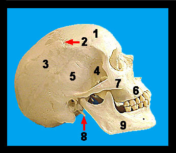
There are two parietal bones, and they form most of the roof and lateral aspects of the cranium. The two parietal bones are joined along the superior midline by the sagittal suture. These bones articulate at the anterior region of the skull with the frontal bone via the coronal suture and at the posterior region of the skull with the occipital bone via the lambdoid suture. Laterally the parietal bones articulate with the temporal bones at the squamous suture. These bones do not contain any distinct surface markings. The temporal bones form a portion of the lateral side of the cranium commonly referred to as the temple. They are situated immediately inferior to the parietal bones and anterior to the occipital bone. The squamous sutures separate the temporal from the parietal bones. At their anterior region the temporal bones articulate with the sphenoid bone. There are three major processes arising from each temporal bone: on the lateral surface the slender zygomatic process of the temporal bone extends to articulate with the zygomatic bone. These two bony structures form the zygomatic arch. On the inferior aspect of the temporal bone two processes arise. Medial and anterior to the mastoid process is the needle-like styloid process. The styloid process serves as an attachment site for muscles of the tongue and neck and for a ligament connecting to the hyoid bone. |
|