Morphology
As a member of the
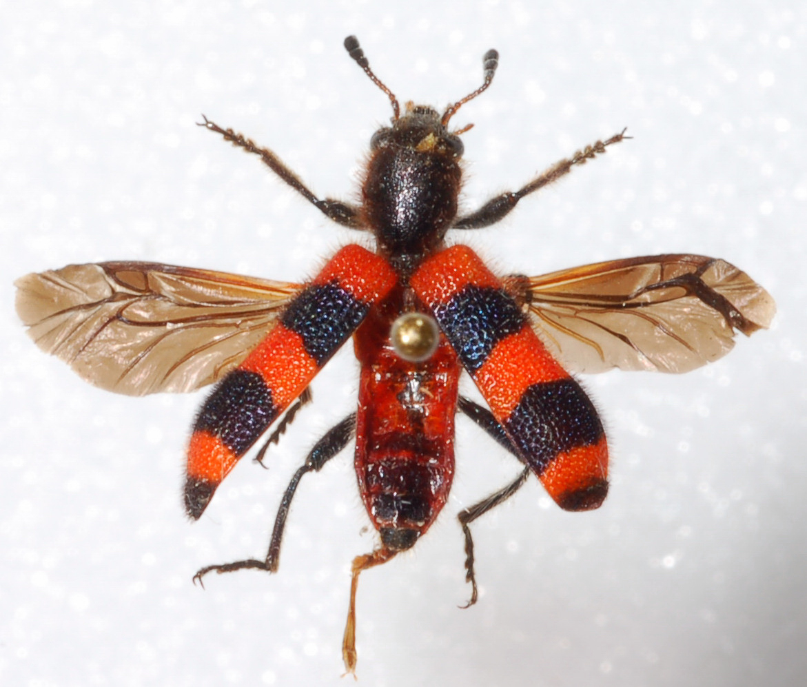
order Coleoptera, Trichodes apivorus has four wings in two
pairs: the elytra and the hind-wings (Burton 1968). The elytra
are a pair of modified forewings which attach at the beetle's
thorax (Jaques 1951), and are hardened to protect the delicate
hind-wings, as well as the otherwise vulnerable soft tissues of
the insect's abdomen
 (Burton 1968). These elytra, or "wing
covers," are part of the exoskeleton protecting the organism (Jaques
1951). The chitonous exoskeleton also covers the head,
thorax and legs and functions as protection from bodily harm and
as an attachment point for bodily tissues, both for locomotion
and body structure (Jaques 1951). This allows the beetle
to maintain its slender and cylindrical shape (Blatchley 1910).
As an attachment point for muscles, the beetle would be unable
to walk or fly without the exoskeletion (Hickman 2009).
The exoskeleton is also responsible for the red and black
coloration of Trichodes apivorus, due to the pigments embedded
within it (Jaques 1951). The outermost cuticle layer
covering the organism also prevents dissication, or water loss
(Hickman et al. 2009).
(Burton 1968). These elytra, or "wing
covers," are part of the exoskeleton protecting the organism (Jaques
1951). The chitonous exoskeleton also covers the head,
thorax and legs and functions as protection from bodily harm and
as an attachment point for bodily tissues, both for locomotion
and body structure (Jaques 1951). This allows the beetle
to maintain its slender and cylindrical shape (Blatchley 1910).
As an attachment point for muscles, the beetle would be unable
to walk or fly without the exoskeletion (Hickman 2009).
The exoskeleton is also responsible for the red and black
coloration of Trichodes apivorus, due to the pigments embedded
within it (Jaques 1951). The outermost cuticle layer
covering the organism also prevents dissication, or water loss
(Hickman et al. 2009).
Each of Trichodes apivorus' three
thoracic segments is equipped with a pair of jointed legs (Blatchley
1910). These limbs attach to the body at ball-and-socket
joints (Jaques 1951). The distal tarsal segments are
covered with setae, providing grip on the surface of a flower,
tree, or any other surface the insect happens to be occupying (Jaques
1951). Though the legs of the adult beetle are long, the
juvenile beetle is equipped with shorter legs to facilitate
movement within the confined spaces of a beehive and find prey (Blatchley
1910).
To navigate its environment and
perceive auditory and
 olfactory
stimuli, a pair of 11-jointed antennae can be found on the
anterior surface of the head (Wolcott 1947). The antennae can also detect taste and
changes in temperature and humidity (Wolcott 1947).
The wide-set compound eyes of Trichodes apivorus can be found on
the anterior surface of the head, and lateral to the antennae
(Dillon 1961). These eyes are made up of many retinal
cells in groups surrounding light receptors, or rhabdom (Wolcott
1947). Because the eyes are incapable of
movement and focus, each lens contributes a small part of an
image, and these images combine to create a field of vision up
to several feet (Wolcott 1947). Several appendages
called maxillae can also be found lateral to the mouth and
mandibles (Jaques 1951). The maxillae are comprised
primarily of maxillary and lateral palpi which serve to hold and
move food to the mandibles for ingestion. The short
alimentary canal common in carnivorous species is well suited to
digesting Trichodes apivorus' choice diet of pollen and insects,
which is high in protein (Jolivet 1998).
olfactory
stimuli, a pair of 11-jointed antennae can be found on the
anterior surface of the head (Wolcott 1947). The antennae can also detect taste and
changes in temperature and humidity (Wolcott 1947).
The wide-set compound eyes of Trichodes apivorus can be found on
the anterior surface of the head, and lateral to the antennae
(Dillon 1961). These eyes are made up of many retinal
cells in groups surrounding light receptors, or rhabdom (Wolcott
1947). Because the eyes are incapable of
movement and focus, each lens contributes a small part of an
image, and these images combine to create a field of vision up
to several feet (Wolcott 1947). Several appendages
called maxillae can also be found lateral to the mouth and
mandibles (Jaques 1951). The maxillae are comprised
primarily of maxillary and lateral palpi which serve to hold and
move food to the mandibles for ingestion. The short
alimentary canal common in carnivorous species is well suited to
digesting Trichodes apivorus' choice diet of pollen and insects,
which is high in protein (Jolivet 1998).
Lastly, the abdomen of Trichodes
apivorus is composed of 10 segments, many of which cannot be
seen externally (Jaques 1951). This seeming absence is due
to modification; the "missing" segments have become structures
within the body. They serve reproductive functions (the
genetalia remain within the body until such a time it is needed)
(Jaques 1951). Each of the visible abdominal segments
bears a pair of respiratory organs called spiracles
(Wolcott 1947), which act as breathing pores to regulate the
size of internal trachae (Jaques 1951). other nutrients
are carried by hemolymph in an open circulatory system powered
by a rudimentary, tube-shaped heart (Wolcott 1947). All
of these adaptations have made Trichodes apivorus
well-suited to the niche it occupies in different stages of its
life cycle.
<<< Life Cycle and Development Home Habitat and Nutrition >>>