Interactions
As
Paragonimus westermani is a parasite, it interacts with
several different species across several different phyla, usually to
that species’ detriment. The dynamic of the interaction is
invariably that of parasite/host. P. westermani has three
major forms, and each form is associated with a different species or
group of species as the host. In order of P. westermani’s
lifecycle, I will list the host species and the parasite’s effect on
them.
First,
the snail....
The first form
of P. westermani is called the miracidia, or ciliated free-living
larval form. Once hatched from its egg, the miracidia swims to its
first host, the freshwater snail. The miracidia infects the snail,
and begins growing into a cercaria (Liu et al. 2008).
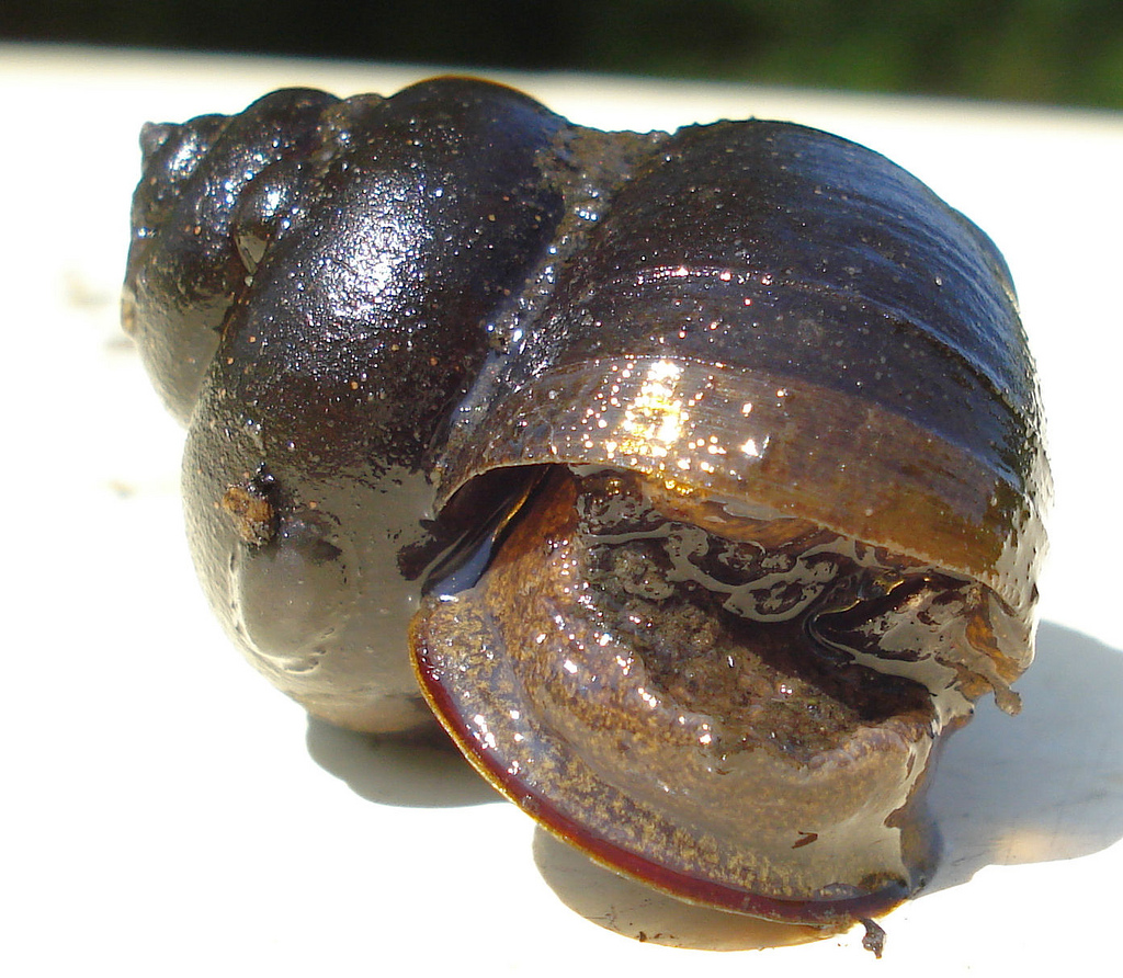 The cercaria
grow inside a group of snail species in the
genus Paludomus. These snails are P. westermani’s
first intermediate host. The cercaria infest the snails’
reproductive organs, and they do so at a rate of approximately .2
percent. (Iwagami et al. 2007). After some time, the
cercariae emerge from the snail, or the snail is eaten by a
predator. Cercariae emergence from an infected snail significantly
increases the snail’s mortality (Koprivnikar et al. 2011).
There are several different second intermediate hosts of P.
westermani, which we will cover next.
The cercaria
grow inside a group of snail species in the
genus Paludomus. These snails are P. westermani’s
first intermediate host. The cercaria infest the snails’
reproductive organs, and they do so at a rate of approximately .2
percent. (Iwagami et al. 2007). After some time, the
cercariae emerge from the snail, or the snail is eaten by a
predator. Cercariae emergence from an infected snail significantly
increases the snail’s mortality (Koprivnikar et al. 2011).
There are several different second intermediate hosts of P.
westermani, which we will cover next.

Then,
the crab.....
After emerging
from or being eaten while inside of their snail host, the cercaria
can “set up shop” in several different organisms. The most common is likely the freshwater crab. In these crabs, the cercaria will encyst
(make a tough layer around) itself while it begins to develop into
an adult fluke. Once encysted, the cercaria is called a
metacercaria. The metacercaria can be found in the crab’s muscles,
hepatopancreas, and gills (Rekha Devi et al. 2012). Another
candidate for the cercaria to infect are crayfish, which led to
human cases of paragonimiasis in the United States after the
crayfish were eaten raw (Lane et al. 2009).

Lastly,
us!!!!
While the first
two stages of P. westermani are certainly troublesome to the
organisms infested with them, we are typically most concerned with
the damage done by the adult version of the fluke. To infest their
definitive host (mammals), the infested second intermediate host
must be eaten raw. The metacercaria may then migrate from the
mammal’s digestive tract to their final destination (mammal lung
tissue). They are able to bore through tissue in this way due to an
enzyme that they secrete. This enzyme dissolves mammalian tissue,
allowing the metacercaria to: migrate, ingest nutrients, and evade
the host’s immune system. Once in place, the metacercaria develop
into adult flukes, which then build a fibrous granuloma (image
right) around
themselves (Na et al. 2005).
Not The
Pinky!!
The metacercaria
may also infest other organs in the mammal’s body, such as the
peritoneum (lining of the intestines) or the brain. There was even
one
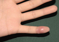 documented case of the fluke that had migrated to the tip of a
patient’s little finger! If in human lung tissue, the fluke will
almost certainly cause symptoms that resemble tuberculosis (TB) or
lung cancer (violent, blood-tinged coughing). Many people with a
P. westermani infestation exhibiting these symptoms are treated
for TB initially. Infestation of nearly any part of the mammal’s
body is considered paragonimiasis. (Sim et al. 2010).
documented case of the fluke that had migrated to the tip of a
patient’s little finger! If in human lung tissue, the fluke will
almost certainly cause symptoms that resemble tuberculosis (TB) or
lung cancer (violent, blood-tinged coughing). Many people with a
P. westermani infestation exhibiting these symptoms are treated
for TB initially. Infestation of nearly any part of the mammal’s
body is considered paragonimiasis. (Sim et al. 2010).
Continue to: Facts