|


| |
|
Mycobacterium
tuberculosis |
|
Characteristics |
|
Mycobacterium tuberculosis is a
rod-shaped (bacillus) bacterium that causes the disease tuberculosis in
humans, as well as other primates, hamsters, dogs, and guinea pigs
(Figure 1).
Since the organism is non-motile, it travels through the air on
particles called droplet nuclei. Droplet nuclei, which range in size
from 1 to 5 μm, are introduced into the air when an infected person
sneezes, coughs, etc. Normal air currents keep the particles airborne
so that they can spread throughout an area. (See the “Pathogenesis”
section for information on how an infection progresses.) In addition to
being non-motile, M. tuberculosis is an obligate aerobe, meaning
that the bacterium can only survive in an environment that contains
oxygen. (See the “Pathogenesis” section to learn more about the
environment in which M. tuberculosis lives.)
|
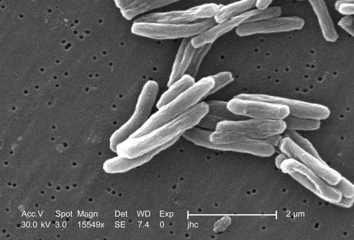
Figure 1. Scanning electron micrograph of Mycobacterium
tuberculosis bacilli.
|
|
Although Mycobacterium tuberculosis
possesses a Gram-positive type cell wall, a cell wall with extensive
peptidoglycan and no outer membrane, the bacterium does not stain with
Gram stain reagents. Gram stain reagents are unable to penetrate the
cell wall of the bacillus because layers of lipids surround the
peptidoglycan in mycobacteria. Unlike most Gram-negative bacteria,
which have a 5-20% lipid content by weight, M. tuberculosis and
other mycobacteria are composed of up to 60% lipids. Many of these
lipids are in the form of mycolic acids. (See the “Adaptations” section
for more on the importance of a high lipid content.)
Since the Gram stain method proves
ineffective on Mycobacterium tuberculosis, acid-fast staining
must be used to make the bacilli visible under a microscope. In the
Ziehl-Neelsen procedure, bacteria from a sputum (mucus coughed up from
the lungs) sample are flooded with a basic solution of carbolfuchsin, a
magenta dye. After heating the slide in a flame, the sample is washed
with water and treated with acid-alcohol to decolorize the bacteria.
Then, a counterstain of methylene blue is applied to the sample. When
the process is complete, M. tuberculosis bacilli appear pink
because they retain the carbolfuchsin during the acid-alcohol
decolorization step (Figure 2). Thus, mycobacteria are classified as acid-fast
bacilli. Bacteria that are not acid-fast appear blue after the
procedure.
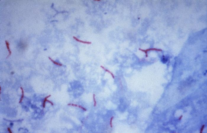
Figure 2. Mycobacterium tuberculosis bacilli stained
using the Ziehl-Neelsen method. |
|