|
Mycobacterium
tuberculosis |
|
Diagnosis |
A tuberculin skin test is
a diagnostic tool for detecting if an individual has been infected with
Mycobacterium tuberculosis. In the United States, the test if
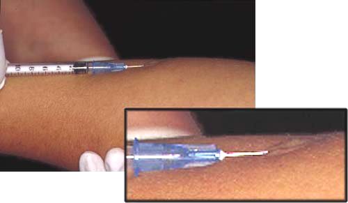
most commonly performed using the Mantoux method. During the procedure,
a tiny amount of purified tuberculin protein derivative (PPD) is
injected just beneath the skin using a needle (Figure 1). If a person
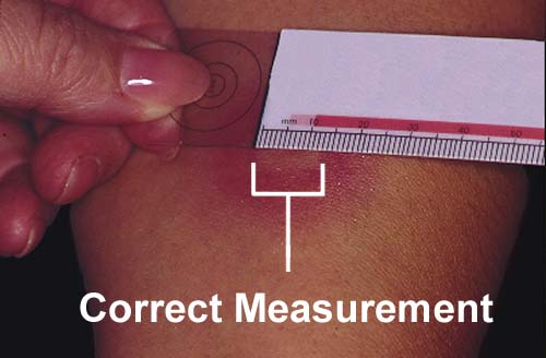
tests positive for tuberculosis, a red, raised bump (induration) will
develop within 48 to 72 hours (Figure 2). The injection site is then
examined by a healthcare professional who decides the next course of
action based on the results. An induration that measures between one
and five millimeters is considered a negative response, so the test is
repeated a week later using a stronger dose of PPD. Sometimes, a false
negative will result if a person was recently infected with the
bacteria, so a tuberculin skin test proves most accurate if performed at
least two to ten weeks after initial exposure. Indurations that range
from five to nine millimeters are considered questionable, so the test
is also repeated a second time in this instance. An induration
measuring 10 to 33 millimeters indicates that a person has been infected
with M. tuberculosis.
|
|
|
 |
|

|
|
A positive skin test does not necessarily mean that an individual has
tuberculosis disease, but rather that an individual has the bacteria
inside of his or her body. Therefore, people with latent tuberculosis
infection and those who have had tuberculosis disease but received
treatment will still test positive. If a positive skin test is
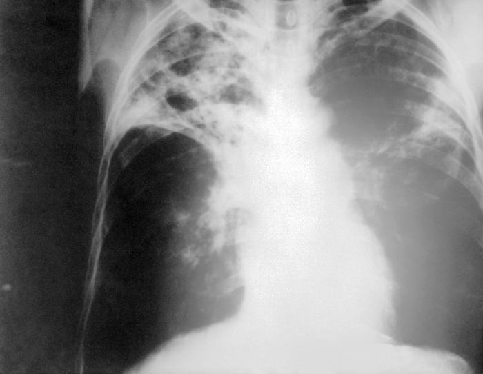
obtained, the healthcare professional will suggest that a chest x-ray be
taken to look for signs of active tuberculosis disease (Figure 3).
Other confirmatory tests include staining and examining sputum samples
or cultivating M. tuberculosis on laboratory media (Figure 4).
Usually, the culture method is utilized only in large hospitals or state
public health facilities because the slow-growing bacterial colonies can
take weeks to appear.
|
|
 |
Figure 3. Chest x-ray of a patient with advanced pulmonary
tuberculosis disease. Calcification of the tubercles allows for
visualization through radiographic imaging. The patient exhibits
the formation of a cavity in the right side of his lungs. Such
cavities result when the interiors of the tubercles become liquefied and
spill out their contents.
|

|