|


| |
|
Mycobacterium
tuberculosis |
|
Pathogenesis |
|
Infection with Mycobacterium tuberculosis
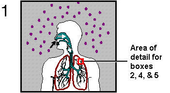
begins when droplet nuclei are inhaled into the upper respiratory tract
through the mouth or the nose (Figure 2, box 1). From the upper respiratory tract, the
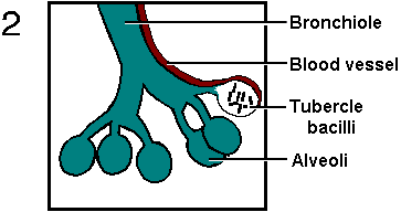
bacilli travel through the bronchi until they reach the alveoli of the
lungs (Figure 2, box 2). Once inside the lungs, alveolar macrophages (immune cells that
engulf foreign particles) ingest the pathogenic organisms, but the mycobacteria do not die. Instead, the bacilli multiply within the
macrophage hosts, causing the macrophages to rupture. The continued
division of M. tuberculosis every 18 to 24 hours attracts more
and more immune cells to the area. In an attempt to control the
infection, some of these cells produce toxic substances that are
supposed to kill the bacilli. The bacilli do not immediately die,
however, so the release of toxic substances also damages the surrounding
lung tissue. When macrophages and other cells of the immune system
encircle this area of dead tissue, the lesion is called a tubercle or
granuloma (Figure 1).
|
|
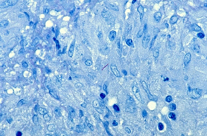 |
Figure 1. Mycobacterium tuberculosis bacilli visible
within granuloma. This tissue sample was taken from the
endometrial layer of the uterus. This is an example of
tuberculosis disease persisting in an area of the body outside of the
lungs.
|
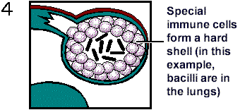
The interior of a tubercle consists of a gelatinous mass of host cells
and bacilli that gives the damaged tissue a cheese-like consistency.
Therefore, this type of tissue death is referred to as caseation necrosis. If the
immune system is successful in preventing the M. tuberculosis
bacilli from multiplying further, the caseous tubercles become
walled-off and calcified (Figure 2, box 4). Although calcified lesions still contain
viable bacteria, the bacteria cannot be spread to other individuals.
When Mycobacterium tuberculosis lies dormant in the lungs, a
person is said to have a latent tuberculosis infection. Such persons,
who represent 85-95% of infected individuals, show no overt symptoms of
disease.
|
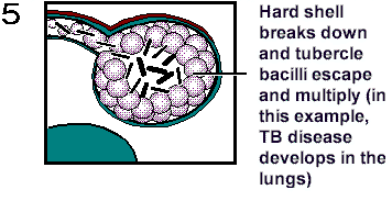
In
5-15% of infected individuals, the immune system fails to prevent the
infection from progressing and the interiors of the caseous lesions become liquefied.
Liquefaction allows viable M. tuberculosis bacilli to spill out
of the tubercles, leaving behind a cavity in the lungs (Figure 2, box 5). When these
bacilli infect lower portions of the lungs or enter the bronchi the
result is an active case of pulmonary tuberculosis disease. People with
pulmonary tuberculosis are capable of spreading the disease to others
through the bacteria in their sputum. They also manifest symptoms such
as weight loss, weakness, night sweats, chest pain, and coughing up
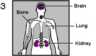
blood. If viable bacilli enter the bloodstream, M. tuberculosis
can travel to organs of the body outside of the lungs (Figure 1, above
and Figure 2, box 3). Known as
extra-pulmonary tuberculosis, this form of the disease is rarely
contagious.
|
 |
 |
|
 |
 |
|
 |
|
As mentioned earlier,
people with latent tuberculosis infection retain viable M.
tuberculosis bacilli within their lungs, even though they are
asymptomatic and not infectious. When a person with latent TB becomes
immunosuppressed because of old age, lifestyle choices, illness, or a
medical condition such as HIV, the dormant bacteria can reactivate
within the calcified tubercles. Thus, people with latent tuberculosis
infection always run the risk of developing active TB disease. |
|