Reproduction
A brief history lesson on Loa
loa.
A French surgeon at San Domingo in
the Caribbean by the name of Mongin, documented the first case
report of the parasite Loa loa, which was published in
1707 when Mongin performed an unsuccessful surgery to remove
the parasitic
 worm from a women’s infected eye (Holmes 2013).
Loa loa was considered benign, or unharmful, until the
late 1980’s and early 1990’s when more cases were being
reported. Despite more and more cases were
reported at this time, the number of cases was still extremely low
resulting in a limited understanding of this disease. To
make matters worse, the list of symptoms associated with
this infectious nematode seems to be almost never ending (Holmes
2013).
worm from a women’s infected eye (Holmes 2013).
Loa loa was considered benign, or unharmful, until the
late 1980’s and early 1990’s when more cases were being
reported. Despite more and more cases were
reported at this time, the number of cases was still extremely low
resulting in a limited understanding of this disease. To
make matters worse, the list of symptoms associated with
this infectious nematode seems to be almost never ending (Holmes
2013).
How does Loa loa get to the
eye in the first place?
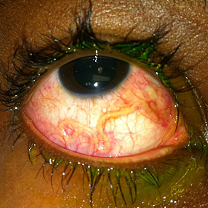
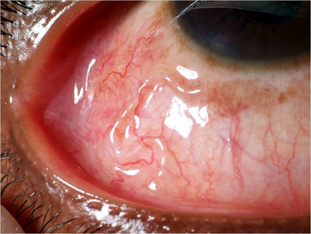 As you can tell by the very graphic
images of human eyes infected by Loa loa, once this worm
grows into an adult, it can live in subcutaneous tissues such as
the
eye. You are probably asking yourself, “But how does it
get to places like your eye in the first place?” Well this is a great
question and I would love to answer it for you!
As you can tell by the very graphic
images of human eyes infected by Loa loa, once this worm
grows into an adult, it can live in subcutaneous tissues such as
the
eye. You are probably asking yourself, “But how does it
get to places like your eye in the first place?” Well this is a great
question and I would love to answer it for you!
Loa loa is a parasitic
species that requires two hosts: An arthropod host commonly known
as the deerfly or the mangofly (both of which are located in the
genus Chrysops) and a
mammalian host (Tyagi et
al. 2011). Loa loa is transferred to its
mammalian host in the form of matured
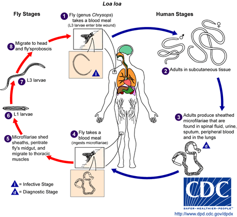 microfilariae, eggs or
larva, when the fly host bites a mammal. Once inside their
new host, the microfilariae travel to the subcutaneous tissues in
the mammal’s eye and remain there until they reach adulthood.
It may take anywhere from one to four years for these
microfilaria to fully mature. Previously, I mentioned that
microfilaria are eggs. Note that eggs is plural as in more
than one! Yes, you read that right, this means that once these eggs
mature there are multiple worms living inside their
mammalian host! Loa loa reproduce sexually where
an adult male and an adult female worm join together to mate
(Roberts and Janovy 2000). They then produce a
new generation of microfilariae through mitosis. These
microfilariae contain protective sheaths that cover each egg in
their first larval stage (Desjardins et al. 2013).
Microfilariae work their way through the mammal’s body by
traveling via the lymphatic system and go on to live in a number
of different places including blood, lungs, urine,
microfilariae, eggs or
larva, when the fly host bites a mammal. Once inside their
new host, the microfilariae travel to the subcutaneous tissues in
the mammal’s eye and remain there until they reach adulthood.
It may take anywhere from one to four years for these
microfilaria to fully mature. Previously, I mentioned that
microfilaria are eggs. Note that eggs is plural as in more
than one! Yes, you read that right, this means that once these eggs
mature there are multiple worms living inside their
mammalian host! Loa loa reproduce sexually where
an adult male and an adult female worm join together to mate
(Roberts and Janovy 2000). They then produce a
new generation of microfilariae through mitosis. These
microfilariae contain protective sheaths that cover each egg in
their first larval stage (Desjardins et al. 2013).
Microfilariae work their way through the mammal’s body by
traveling via the lymphatic system and go on to live in a number
of different places including blood, lungs, urine,
.jpg) sputum, and
even spinal fluid! When a new fly bites the infected
mammal and takes a blood contaminated with microfilariae, it
becomes the new
arthropod host. Once ingested,
microfilaria lose their protective sheaths and are able to
migrate from the stomach or gut of their new host to the
thoracic muscles where they undergo three larval developmental
stages (Desjardins et al. 2013). Then they return to the mouth of the fly upon the third
developmental stage. Once the matured microfilariae make
their way to the fly’s mouth, they wait for the fly to bite a
mammal which will become the new host for young Loa loa.
Thus the cycle repeats itself.
sputum, and
even spinal fluid! When a new fly bites the infected
mammal and takes a blood contaminated with microfilariae, it
becomes the new
arthropod host. Once ingested,
microfilaria lose their protective sheaths and are able to
migrate from the stomach or gut of their new host to the
thoracic muscles where they undergo three larval developmental
stages (Desjardins et al. 2013). Then they return to the mouth of the fly upon the third
developmental stage. Once the matured microfilariae make
their way to the fly’s mouth, they wait for the fly to bite a
mammal which will become the new host for young Loa loa.
Thus the cycle repeats itself.
If you would like to learn more about
the relationships between Loa loa and its two hosts, we
encourage you to check out our INTERACTIONS
page!
Return to HOME
Return to HOME